
Nucleoside based proliferation imaging markers

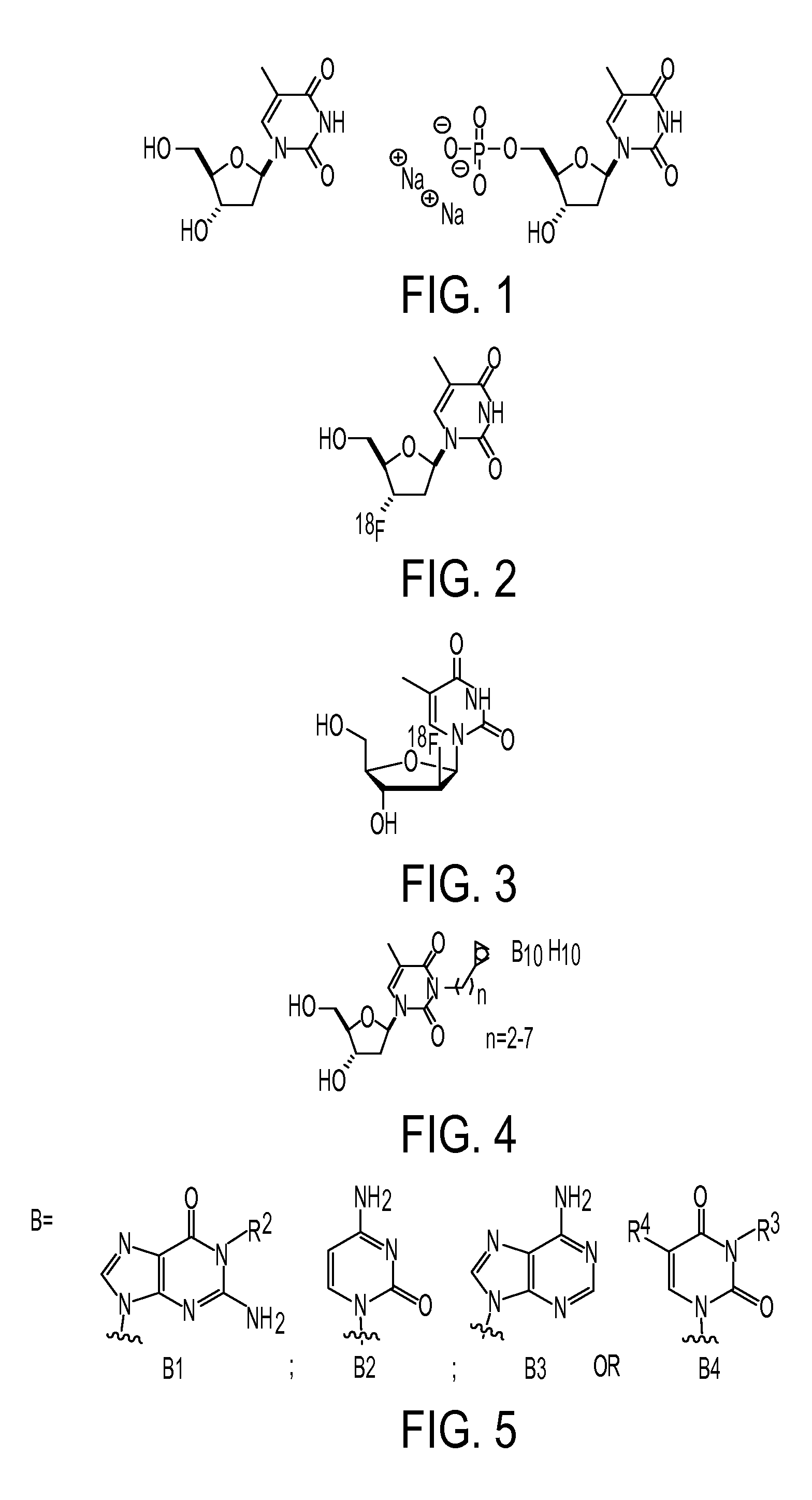
a proliferation imaging and nucleoside technology, applied in the field of nucleoside based proliferation imaging markers, can solve the problems of limited limited specificity of fdg-pet, and accumulation in inflammatory tissue, so as to improve the sensitivity of pet for tumor response prediction, improve the pharmacokinetic profile, and improve the signal to noise ratio

Inactive Publication Date: 2011-04-19
SIEMENS MEDICAL SOLUTIONS USA INC
View PDF16 Cites 7 Cited by
  • Summary
  • Abstract
  • Description
  • Claims
  • Application Information

AI Technical Summary

Benefits of technology

The patent text describes new compounds that can be used to detect and image cell proliferation in the body using positron emission tomography (PET) imaging. These compounds have improved pharmacokinetic profiles, meaning they can be better absorbed and retained by cancer cells, leading to stronger signals and better sensitivity. The compounds have higher rates of phosphorylation relative to another molecule called FLT, which is important for cell proliferation. The compounds can also be used as surrogate markers to monitor the effectiveness of therapy for cancer. Overall, this technology can help clinicians better stage tumors, predict treatment response, and monitor therapy effectiveness.

Problems solved by technology

While the clinical use of PET for detecting cancer is growing, FDG based imaging does have limitations.
Accumulation in inflammatory tissue limits the specificity of FDG-PET.
Conversely, nonspecific FDG uptake may also limit the sensitivity of PET for tumor response prediction.
Further, physiological high normal background activity (i.e., in the brain) can render the quantification of cancer-related FDG-uptake impossible in some areas of the body.
Non-radioactive FLT was originally developed as an antiviral then as a potential therapeutic for HIV; however, its toxicity profile prevented its therapeutic use.
This low tumor uptake has made imaging difficult because of the low signal to noise ratios.
Additionally, the high hepatic retention of FLT prevents accurate imaging of tumors and metastases near the liver thus limiting the utility of FLT in the clinical setting.
While its uptake in cancer cells appears promising, the multistep radiosynthetic method for preparing this molecule severely limits its use as a standard clinical biomarker.

Method used


Image

  • Nucleoside based proliferation imaging markers
  • Nucleoside based proliferation imaging markers
  • Nucleoside based proliferation imaging markers

Examples


examples

[0079]General. The human Thymidine Kinase-1 was ordered from Biaffin GmbH & Co KG. The LC / MS analyses were performed on an Agilent 1100 series LC / MSD (SL) using a 30×2.1 mm Zorbax C8 column with a Phenomenex C18 pre-column. Compound detection was accomplished by electrospray mass spectroscopy in positive selected ion mode (LC / MS-SIM). The elution solvents, acetonitrile and water, contained 0.05% TFA. Nuclear magnetic resonance (NMR) spectra were obtained on a Bruker AMX 300 MHz spectrometer. 19F NMR spectra were recorded on a Bruker AMX 282.35 MHz spectrometer. The mass spectra were recorded on an Agilent 1100 series LC / MSD with electrospray mass spectroscopic detection. Flash column chromatography was performed either on Merck silica gel (40-63 μm) using the solvent system indicated or on a CombiFlash purification system on silica gel cartridges. The radiochemical and chemical purities were analyzed by RP-HPLC.

Synthesis of Precursors and Standards:

[0080]

[0081]Synthesis of 2: To a r...


PUM

PropertyMeasurementUnit
molecular weightaaaaaaaaaa
temperatureaaaaaaaaaa

Abstract

Disclosed herein are novel radiolabeled nucleosides and methods for detecting cellular proliferation in a mammal, the method comprising administrating an effective amount of a radiolabeled nucleoside; the method comprising: a) administering to the mammal a diagnostically effective amount of the nucleoside to the mammal; b) allowing the nucleoside to distribute into the effective tissue; and c) imaging the tissue, wherein an increase in binding of the compound to tissue compared to a normal control level of binding indicates that the mammal is suffering from a disease involving cellular proliferation.

Description

CROSS-REFERENCE TO RELATED APPLICATIONS[0001]This application claims priority to U.S. Provisional Application No. 60 / 904,223, filed Mar. 1, 2007.[0002]The present invention relates to radioactively labeled nucleoside analogs substituted at the 3-N, 3′-O and 2′-O positions with various linkers useful for modifying their in vivo pharmacokinetics. In one embodiment, the nucleoside analogs are thymidine analogs. These compounds display high cellular uptake and phosphorylation rates, undergo facile radiolabeling and display favorable tumor to background uptake ratios. Preferred compounds localize within tumors having high populations of cells in the S-phase. Preferred compounds can be used to detect rapidly proliferating cells in vivo and thus identify patients with diseases marked by abnormally proliferating cells such as cancer. Additionally, these analogs may be used as surrogate markers to monitor efficacy of therapy.BACKGROUND OF THE INVENTION[0003]PET, being a molecular imaging tec...

Claims


Application Information

Patent Timeline
Patent Type & Authority Patents(United States)
IPC IPC(8): C07H21/02C07H21/04
CPCA61K51/0491C07H19/073C07H19/067C07H19/06
Inventor KOLB, HARTMUTH C.WALSH, JOSEPH C.YEH, ROBERT MCHEN, KAIGANGADHARMATH, UMESHDUCLOS, BRIANMOCHARLA, VANI P.KARIMI, FARHADPADGETT, HENRY C.LIANG, QIANWAZHAO, TIEMING
Owner SIEMENS MEDICAL SOLUTIONS USA INC
Learn More