Production of papillomavirus capsid protein and virus-like particles

a technology of capsid protein and papillomavirus, which is applied in the field of papillomavirus, can solve the problems of limiting the usefulness of the study, difficult to conduct the study, and high cost of the system

Active Publication Date: 2011-11-22
UNIVERSITY OF ROCHESTER
View PDF33 Cites 9 Cited by
  • Summary
  • Abstract
  • Description
  • Claims
  • Application Information

AI Technical Summary

Benefits of technology

[0015]Virus-like particle(s) produced in accordance with the invention, can be used in diagnostic assays, can play a role in the identification and characterization of an HPV cell receptor, and can be used for vaccine development (both therapeutic and prophylactic). It is understood that the method of the invention as described herein for pro...

Problems solved by technology

However, difficulties in propagating HPV in vitro has led to the development of alternative approaches to antigen production for immunologic studies.
These studies have been difficult to conduct because most naturally occurring HPV-induced lesions produce few particles.
Although this system is capable of producing quantities of infectious virus that could be used for the development of a serologic test for genital HPV infection, this system is very expensive and cumbersome.
Furthermore, only one genital HPV type has so far been propagated in this system, thus, limiting its usefulness.
In addition, the infectious virus produced using this system represents a biohazard and, therefore, would be difficult to use in a vaccine formulation.
However, the authors were not able to produce VLPs with a vector expressing L1 alone.
However, Minson fails to disclose the expression of the L1 protein or production of HPV virus-like particles (VLPs).
However, the inventors fail to disclose any sequences directed to the major late open reading frame, L1.
However, the inventors do not disclose infected animals that produced antibodies which recognize this sequence.
In addition, the amino acid sequences disclosed (10 amino acids) a...

Method used


Image

  • Production of papillomavirus capsid protein and virus-like particles
  • Production of papillomavirus capsid protein and virus-like particles
  • Production of papillomavirus capsid protein and virus-like particles

Examples


example i

Materials and Methods

1. HPV-11 Viral DNA And pVL11L1 Baculovirus Transfer Vector Construction

[0039]HPV-11 genomic DNA was obtained from virus particles which were purified from experimentally induced athymic mouse xenografts as described by Rose et al., Expression of the full-length products of the HPV-6b and HPV-11 L2 open reading frames by recombinant baculovirus, and antigenic comparisons with HPV-11 whole virus particles, 1990, J. Gen. Virol., vol. 71, pp. 2725-2729, which disclosure is hereby incorporated by reference. The L1 coding sequence was cloned by PCR amplification of purified genomic DNA, using primers designed to introduce BglII and EcoRI restriction enzyme sites at the 5′ and 3′ ends, respectively. The forward and reverse primer sequences, respectively, were, 5′-CGC AGA TCT ATG TDG CCT AGC-3′ (SEQ. ID. NO. 1) and 5′-CAT ATG AAT TCC CAC AAC ACA CTG ACA CAC-3′(SEQ. ID. NO. 2). Restriction sites (underlined) were introduced proximal to the putative L1 start codon (bold ...

example ii

Expression And Immunologic Detection Of Recombinant HPV-11 L1 Protein In Sf-9 cells

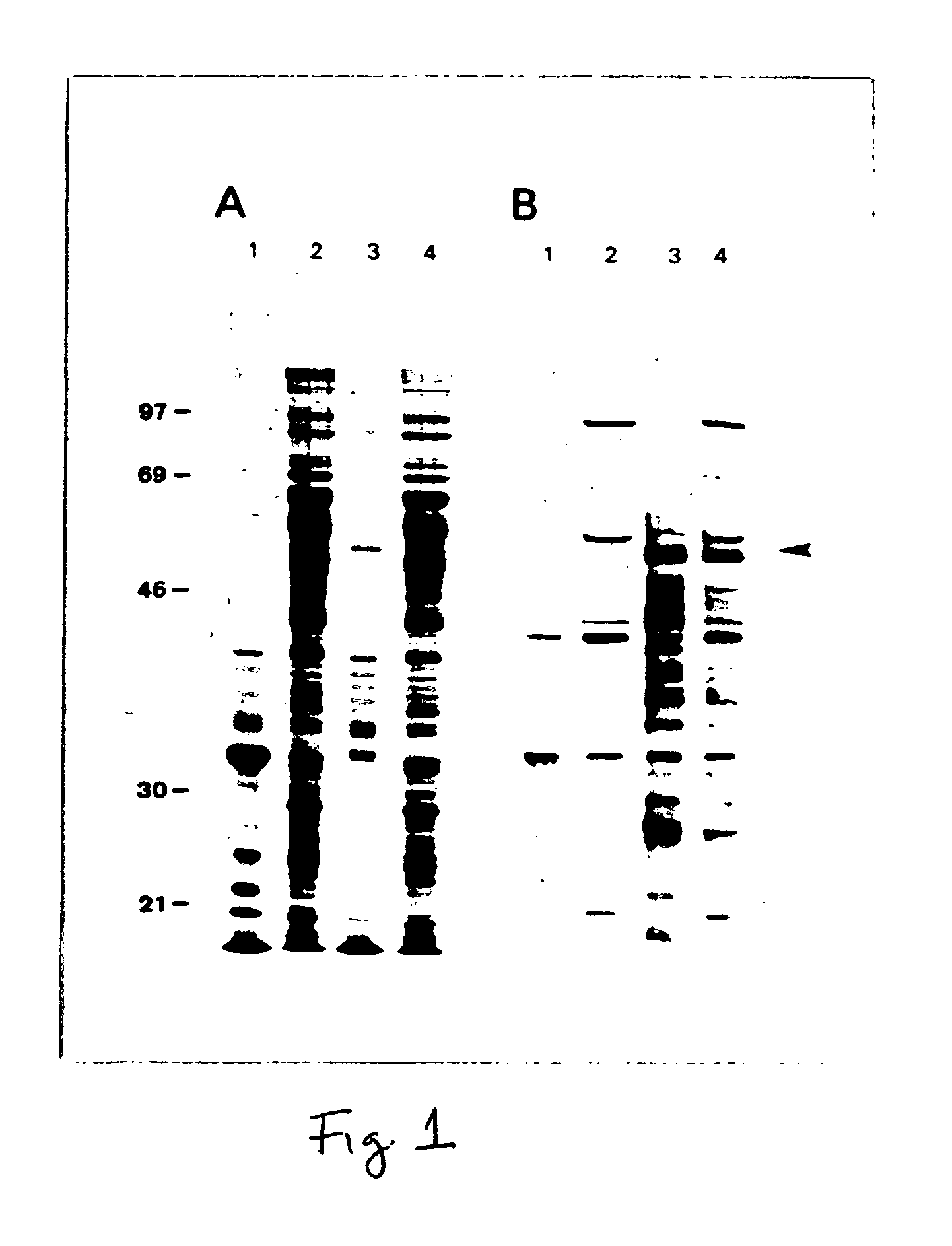
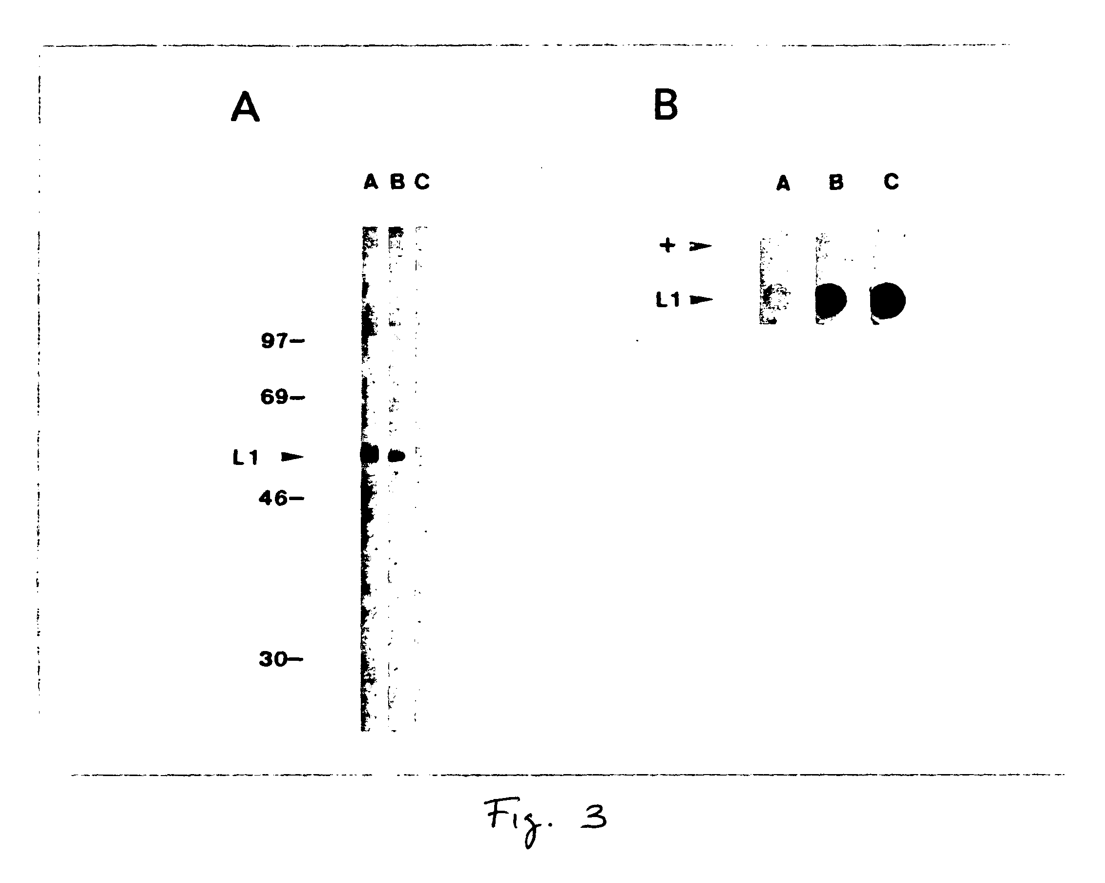
[0048]SDS-PAGE analysis of total Sf-9 cell proteins from insect cells infected with the recombinant virus Ac11L1 demonstrated a novel 55 kD protein seen by Coomassie-blue staining in Ac11L1-infected cells (FIG. 1A, lane 3). With reference to FIGS. 1 (A and B), FIG. 1A shows Coomassie-stained SDS polyacrylamide gel of wild-type AcNPV and recombinant Ac11L1-infected Sf-9 cell lysates and FIG. 1B shows Western blot of wild-type AcNPV and recombinant Ac11L1-infected Sf-9 cell lysates probed with a rabbit polyclonal antiserum specific for the HPV L1 common epitope. Non-recombinant (lanes 1,2) and recombinant L1-infected (lanes 3,4) Sf-9 cell lysates were fractionated into insoluble (lanes 1,3) and soluble (lanes 2,4) fractions, and electrophoresed on 10% polyacrylamide gels. Molecular reference (Mr) markers are displayed at the left, and the arrow at the right indicates the approximate position of recombin...

example iii

Electron Microscopic Visualization of VLPs

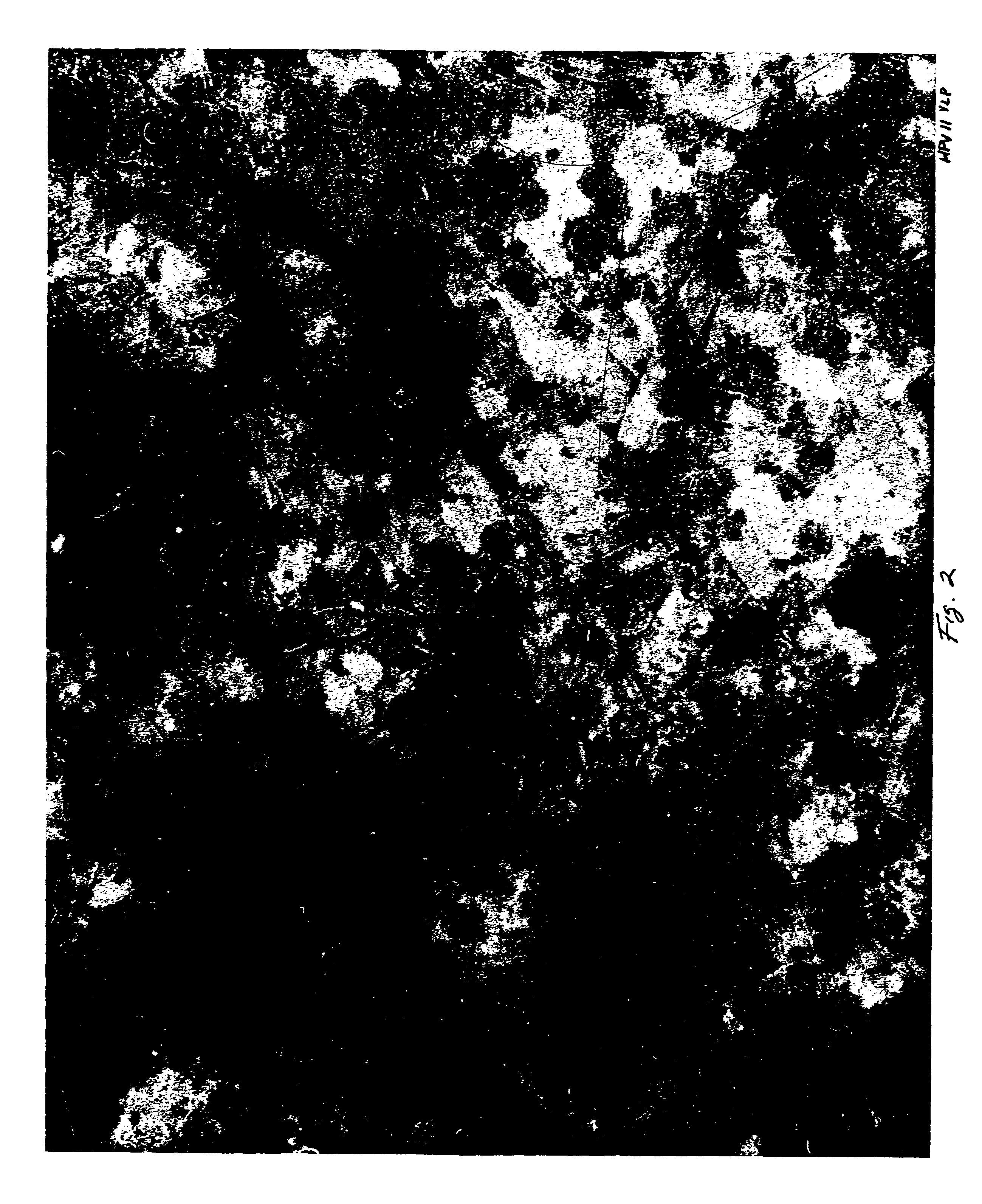
[0049]Electron micrographs of negatively stained preparations of sucrose banded VLPs (FIGS. 2, 6, and 7) showed distinct VLPs. FIG. 2 shows HPV-11 capsid-like particles which were present at the 50-60% interface of the sucrose density gradient. FIG. 6 shows HPV type 6b (HPV-6b) capsid-like particles which resulted from the expression of the HPV-6b L1 coding sequence in the baculovirus system, and which were purified in exactly the same manner. FIG. 7 demonstrates that this method is also suitable for the production of HPV type 16 (HPV-16) VLPs, upon expression of the HPV-16 L1 coding sequence. Particle diameters determined by direct measurement of the VLPs in FIG. 2, were approximately 52 nm. This measurement is consistent with the diameter of isolated papillomavirus virions as described by Klug et al., Structure of viruses of the papilloma-polyoma type I: Human wart virus, 1965, J. Mol. Biol., vol. 11, pp. 403-423, which disclosure is hereb...


PUM

PropertyMeasurementUnit
Diameteraaaaaaaaaa
Volumeaaaaaaaaaa
Sizeaaaaaaaaaa

Abstract

The present invention is directed to a method of expressing the papillomavirus capsid protein coding sequence in a cell using an expression system under conditions facilitating expression of the protein in the cell.
In another aspect of the invention, it has been discovered that virus-like particle(s) (VLPs), fragment(s), capsomer(s) or portion(s) thereof are formed from the papillomavirus capsid protein. It was further discovered that the virus-like particle(s) comprises antigenic characteristics similar to those of native infectious papillomavirus particles.
In an embodiment of the invention, there is provided a method of expressing the L1 major capsid protein of human papillomavirus type-11 (HPV-11) in Sf-9 insect cells using the baculovirus expression system, and the production of HPV-11 virus-like particles.

Description

FIELD OF THE INVENTION[0001]The present invention relates generally to papillomavirus (PV). More particularly, the invention relates to a method of expressing the human papillomavirus (HPV) capsid protein coding sequence using the baculovirus expression system, production of HPV virus-like particles (VLPs) and use of these VLPs in production of antibodies which recognize epitopes on HPV, and for HPV vaccine development, and for development of serologic tests for the detection of HPV infection.BACKGROUND OF THE INVENTION[0002]The family Papovaviridae constitutes a group of DNA viruses that induce both lytic infections and either benign or malignant tumors. Structurally, all are naked icosahedral virions with 72 capsomeres and contain double-stranded circular DNA. Viruses included in the family are: (1) human and animal papillomaviruses, (2) mouse polyomavirus, (3) simian vacuolating virus, and (4) human viruses BK and JC.[0003]Human papillomaviruses (HPV) infect cutaneous, genital, o...

Claims


Application Information

Patent Timeline
IPC IPC(8): A61K39/12
CPCA61K39/12A61K2039/5258C12N2710/20034C12N2710/20023C12N2710/20022
Inventor ROSE, ROBERTBONNEZ, WILLIAMREICHMAN, RICHARD
Owner UNIVERSITY OF ROCHESTER
Why Eureka
Try Eureka