
Methods of using adipose derived stem cells to promote wound healing

a technology of regenerative cells and stem cells, applied in the field of regenerative cells, can solve the problems of slowing down the body's own healing process, significant individual morbidity, and profound reduction in quality of life, and achieve the effect of promoting differentiation

Inactive Publication Date: 2012-01-31
LOREM VASCULAR PTE LTD
View PDF150 Cites 59 Cited by
  • Summary
  • Abstract
  • Description
  • Claims
  • Application Information

AI Technical Summary

Problems solved by technology

Approximately five million Americans suffer from chronic open sores often due to limited blood flow which can slow the body's own healing process.
Inadequate and / or incomplete wound healing, however, can lead to significant individual morbidity including a profound reduction in quality of life.
Sores can become seriously infected, gangrenous and in some cases require amputation.
For example, the primary cause of non-traumatic amputation due to inadequate wound healing is diabetes.
More than 67,000 patients with diabetes require surgical amputation each year, necessitating long and costly rehabilitation.
For many, mobility and independence are severely affected, permanently altering their quality of life.
At its extreme, poor wound healing can serve as the root cause of death.
For patients who have chronic wounds that are difficult to heal, basic medical and surgical care is essential, but not always enough.
Broad application of regenerative cellular therapy, however, has been hindered by questions of cellular retention and the high costs associated with extensive ex vivo manipulation.
However, although stem cell populations have been shown to be present in one or more of bone marrow, skin, muscle, liver and brain (Jiang et al., 2002b; Alison, 1998; Crosby and Strain, 2001), their frequency in these tissues is low.
Although cell culture steps may provide increased cell number, purity, and maturity, they do so at a cost.
This cost can include one or more of the following technical difficulties: loss of cell function due to cell aging, loss of potentially useful non-stem cell populations, delays in potential application of cells to patients, increased monetary cost, and increased risk of contamination of cells with environmental microorganisms during culture.

Method used


Image

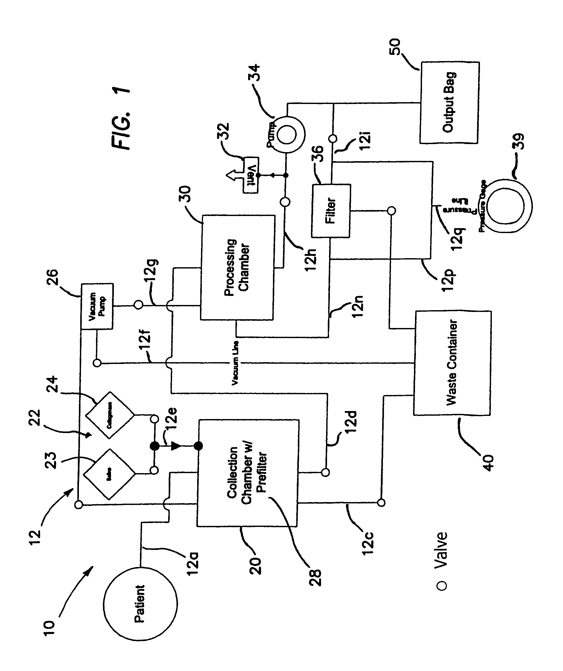
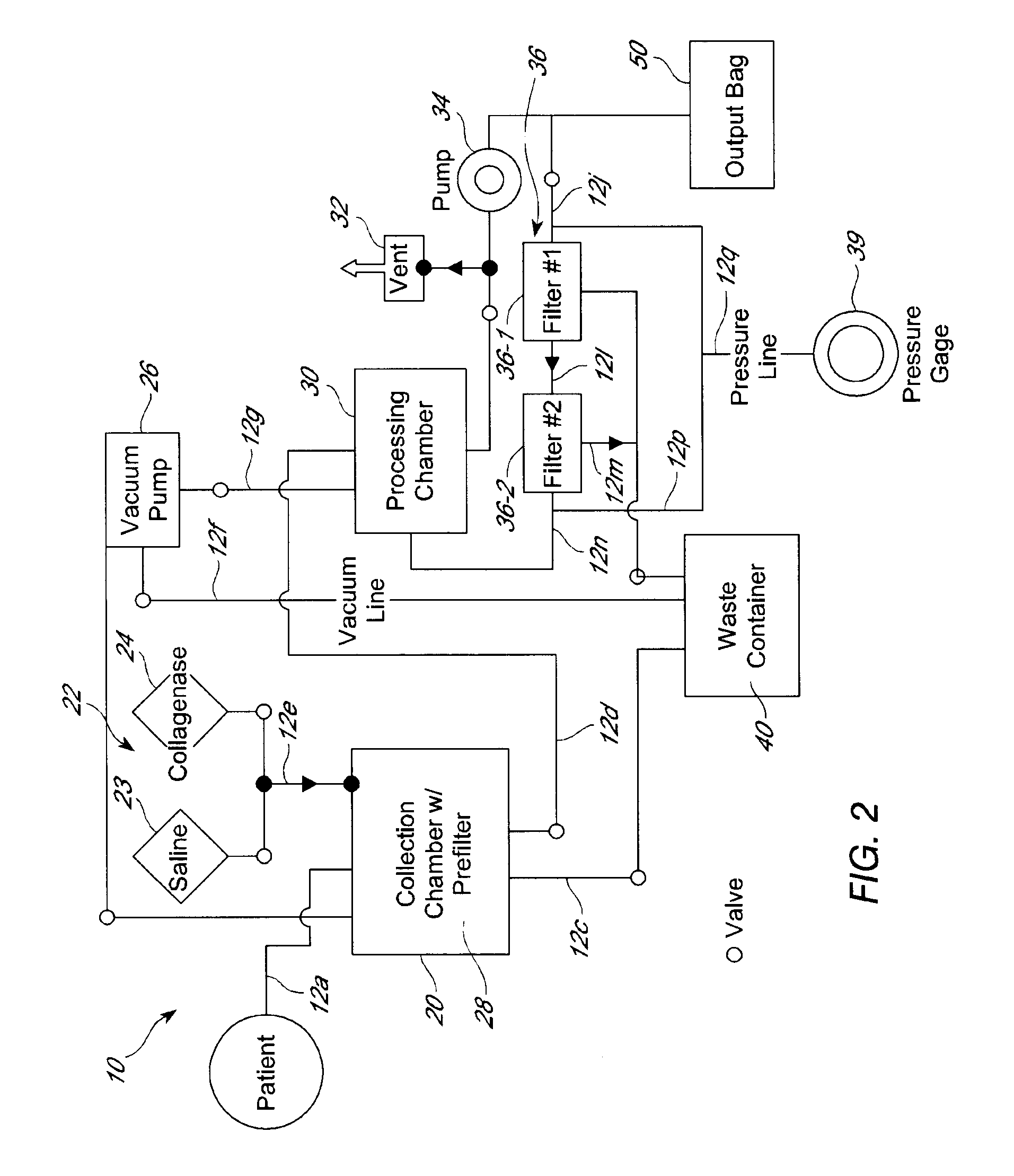
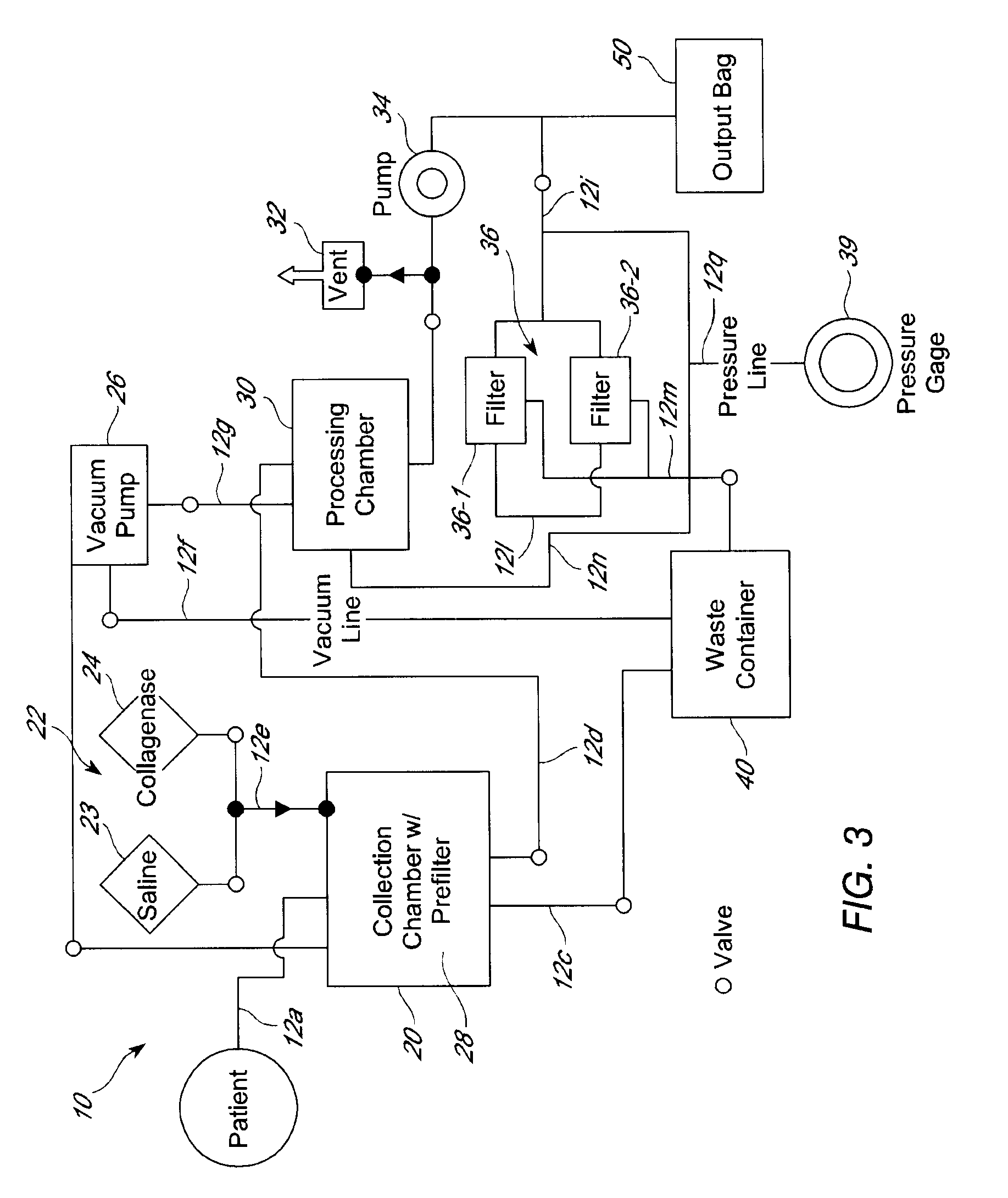
  • Methods of using adipose derived stem cells to promote wound healing
  • Methods of using adipose derived stem cells to promote wound healing
  • Methods of using adipose derived stem cells to promote wound healing

Examples


example 1

Expression of Angiogenic Growth Factor, VEGF, by Regenerative Cells

[0193]Vascular Endothelial Growth Factor (VEGF) is one of the key regulators of angiogenesis (Nagy et al., 2003; Folkman, 1995). Placenta Growth Factor, another member of the VEGF family, plays a similar role in both angiogenesis as well as arteriogenesis. Specifically, transplant of wild-type (PIGF + / +) cells into a PIGF knockout mouse restores ability to induce rapid recovery from hind limb ischemia (Scholz et al., 2003).

[0194]Given the importance of angiogenesis and arteriogenesis to the revascularization process, PIGF and VEGF expression by the regenerative cells of the present invention was examined using an ELISA assay (R&D Systems, Minneapolis, Minn.) using adipose derived regenerative cells from three donors. One donor had a history of hyperglycemia and Type 2 diabetes (a condition highly associated with microvascular and macrovascular disease). Regenerative cells from each donor were plated at 1,000 cells / cm...

example 2

Regenerative Cells Contain Cell Populations That Participate in Angiogenesis

[0197]Endothelial cells and their precursors, endothelial progenitor cells (EPCs), are known to participate in angiogenesis. To determine whether EPCs are present in adipose derived regenerative cells, human adipose derived regenerative cells were evaluated for EPC cell surface markers, e.g., CD-34.

[0198]ADCs were isolated by enzymatic digestion of human subcutaneous adipose tissue. ADCs (100 ul) were incubated in phosphate saline buffer (PBS) containing 0.2% fetal bovine serum (FBS), and incubated for 20 to 30 minutes at 4° C. with fluorescently labeled antibodies directed towards the human endothelial markers CD-31 (differentiated endothelial cell marker) and CD-34 (EPC marker), as well as human ABCG2 (ATP binding cassette transporter), which is selectively expressed on multipotent cells. After washing, cells were analyzed on a FACSAria Sorter (Beckton Dickenson—Immunocytometry). Data acquisition and analy...

example 3

In Vitro Development of Vascular Structures in Regenerative Cells

[0201]An art-recognized assay for angiogenesis is one in which endothelial cells grown on a feeder layer of fibroblasts develop a complex network of CD31 -positive tubes reminiscent of a nascent capillary network (Donovan et al., 2001). Since adipose derived regenerative cells contain endothelial cells, EPCs and other stromal cell precursors, we tested the ability of these regenerative cells to form capillary-like structures in the absence of a feeder layer. Regenerative cells obtained from inguinal fat pads of normal mice developed capillary networks two weeks after culture (FIG. 18A). Notably, regenerative cells from hyperglycemic mice with streptozotocin (STZ)-induced Type I diabetes eight weeks following administration of STZ formed equivalent capillary networks as those formed by cells from normal mice (FIG. 18B).

[0202]In a subsequent study, adipose derived regenerative cells were cultured in complete culture medi...


PUM


Abstract

Cells present in adipose tissue are used to promote wound healing in a patient. Methods of treating patients include processing adipose tissue to deliver a concentrated amount of regenerative cells obtained from the adipose tissue to a patient. The methods may be practiced in a closed system so that the regenerative cells are not exposed to an external environment prior to being administered to a patient. Accordingly, in a preferred method, cells present in adipose tissue are placed directly into a recipient along with such additives necessary to promote, engender or support a therapeutic benefit.

Description

RELATED APPLICATIONS[0001]This application is a continuation-in-part application of U.S. application Ser. No. 10 / 316,127, filed on Dec. 9, 2002 now abandoned, entitled SYSTEMS AND METHODS FOR TREATING PATIENTS WITH PROCESSED LIPOASPIRATE CELLS, which claims the benefit of U.S. Provisional Application No. 60 / 338,856, filed Dec. 7, 2001. The contents of the aforementioned applications are expressly incorporated herein by this reference.BACKGROUND OF THE INVENTION[0002]1. Field of the Invention[0003]This invention generally relates to regenerative cells derived from a wide variety of tissues, and more particularly, to adipose-derived regenerative cells (e.g., stem and / or progenitor cells), methods of using adipose-derived regenerative cells, compositions containing adipose-derived regenerative cells, and systems for preparing and using adipose-derived regenerative cells which are used to promote wound healing, e.g., wounds resulting from diabetes, chronic peripheral vascular disease an...

Claims


Application Information

Patent Timeline
Patent Type & Authority Patents(United States)
IPC IPC(8): C12N5/00A61M1/00A61K31/436A61K35/02A61K35/12A61K35/28A61K35/36A61K35/44A61K38/00A61K38/16A61K45/00A61L27/00A61P1/00A61P1/16A61P3/00A61P3/10A61P9/10A61P11/00A61P13/12A61P17/00A61P19/00A61P19/02A61P21/00A61P25/00A61P27/02A61P37/06C12M3/06C12N5/0775
CPCA61K35/12C12N5/069A61K35/28A61K35/36A61K35/44C12N5/0667A61L27/54A61K9/0014A61K35/35A61L15/40A61L15/44A61L27/3604A61K38/1866A61K45/06A61L27/3834A61L27/56A61L2300/64C12N2506/1384A61L2300/412A61K2300/00A61P1/00A61P1/16A61P3/00A61P3/10A61P9/00A61P9/10A61P11/00A61P13/12A61P17/00A61P17/02A61P19/00A61P19/02A61P21/00A61P25/00A61P27/02A61P37/06
Inventor FRASER, JOHN K.HEDRICK, MARC H.DANIELS, ERIC
Owner LOREM VASCULAR PTE LTD
Learn More