Compositions and methods for treating cartilage disease

a cartilage disease and cartilage technology, applied in the direction of drug compositions, skeletal/connective tissue cells, prostheses, etc., can solve the problems of virtually no potential for self-repair, inadequate repair of cartilage defects, and lack of vascular and neural networks of articular cartilage, so as to improve the lubricating function of joints, promote effective cartilage regeneration, and improve the effect of lubricating function

Active Publication Date: 2013-02-12
MOCHIDA PHARM CO LTD +1
View PDF17 Cites 5 Cited by
  • Summary
  • Abstract
  • Description
  • Claims
  • Application Information

AI Technical Summary

Benefits of technology

[0008]For example, Reference 1 discloses an alginate gel comprising a mixture of a soluble alginate and an insoluble alginate / gel, while References 2, 3 and 4 disclose the use of alginate beads. In Reference 2, alginic acid can be used as a carrier that does not impart any disadvantageous effects on an injured site, although alginic acid itself is discussed as not having any therapeutic effects. In addition, Reference 4 discloses that chondrocytes embedded in alginate beads were not observed to fuse to host tissue after transplanting to a rabbit cartilage defect. In addition, although alginate beads are required to be applied by being pressed into a defect, since it is necessary to produce beads that match the size of the defect, their use in the actual clinical setting is technically difficult. Reference 5 discloses a graft in which chondrocytes are suspended in sodium alginate and injected into a rabbit cartilage defect followed by curing the surface with CaCl2 solution, wherein although normal cartilage tissue is formed, fibrous cartilage is formed in the case of applying only alginic acid to the cartilage defect without containing cells.
[0009]In addition, research is progressing on the use of collagen sponge and the like as a cell scaffold as an example of attempts to use mesenchymal stem cells for cartilage regenerative therapy. Although methods involving transplantation of mesenchymal stem cells following in vitro differentiation to chondrocytes, and methods involving transplantation of mesenchymal stem cells without differentiating have been considered, there is still continuing debate over which utilization method is optimal (Reference 6).
[0010]Since cartilage defects in osteoarthritis (OA) occur over a wide range and in regions subjected to loads, their repair by transplant or regenerative therapy is considered to be difficult. Those cartilage defects eligible for cartilage regeneration by cell transplant as described above are limited to partial cartilage defects caused mainly by sporting activities or trauma. Treatment of osteoarthritis focuses primarily on the removal of pain and inflammation at the affected area, and is commonly treated overseas with administration of non-steroid anti-inflammatory drugs. However, since renal function may be depressed in elderly patients, continuous oral administration of non-steroid anti-inflammatory drugs may be difficult from the viewpoint of safety. Products incorporating hyaluronic acid, which is a component of cartilage synovial fluid, improve the lubricating function of joints by being administered into a joint, and since these products also having analgesic action, they are widely used as joint function improving agents for osteoarthritis. However, since there is ultimately no other choice than to replace the joint with an artificial joint in severe cases of osteoarthritis in which joint damage has progressed, there is a desire for the development of a novel therapeutic drug.REFERENCES
[0012]2. Cay M. Mierisch et al., “Transforming Growth Factor β in Calcium Alginate Beads for the Treatment of Articular Cartilage Defects in the Rabbit”, The Journal of Arthroscopic and Related Surgery, Vol. 18, No. 8 (October), 2002: pp. 892-900
[0013]3. David R. Diduch et al., “Marrow Stromal Cells Embedded in Alginate for Repair of Osteochondral Defects”, The Journal of Arthroscopic and Related Surgery, Vol. 16, No. 6 (September), 2000: pp. 571-577
[0014]4. David R. Diduch et al., “Chondrocyte Transplantation into Articular Cartilage Defects with Use of Calcium Alginate: The Fate of the Cells”, J Bone Joint Surg. Am. 85: 2003, pp. 1757-1767

Problems solved by technology

However, articular cartilage lacks vascular and neural networks.
Consequently, it has virtually no potential for self-repair, and in the case of the formation of large cartilage defects in particular, the cartilage defect is not adequately repaired.
Consequently, when a cartilage defect is formed, joint pain and loss of function are brought about that frequently progress to osteoarthritis.
In addition, a cartilage defect can reach over a broad range as a result of symptoms progressing from the initial stages of osteoarthritis that began with wear of the surface of articular cartilage due to aging or excessive joint usage.
In this manner, since articular cartilage lacks an adequate self-repair ability, surgical procedures are required to treat cartilage injuries, examples of which include mosaicplasty, microfracture, drilling, abrasion and debridement.
However, these techniques have limitations with respect to cartilage defects covering a wide area, and cartilage regenerated by these methods is in the form of fibrous cartilage having different mechanical properties than hyaline cartilage.
However, recent clinical studies have reported that ACI does not yield significantly superior results when compared with other techniques for repair of articular cartilage defects.
The first is the technical difficulty associated with fixing the cells and scaffold to the cartilage defect and covering the defect with a periosteal flap.
Moreover, several complications associated with the periosteal flap have been reported, including periosteal hypertrophy, defect formation and intra-articular adhesion.
The other reason involves limitations on the use of chondrocytes.
Another problem is that although ACI requires cartilage to be harvested from a non-weight-bearing site of the joint, donor sites remain problematic since chondrocytes are harvested there from.
In addition, although alginate beads are required to be applied by being pressed into a defect, since it is necessary to produce beads that match the size of the defect, their use in the actual clinical setting is technically difficult.
Since cartilage defects in osteoarthritis (OA) occur over a wide range and in regions subjected to loads, their repair by transplant or regenerative therapy is considered to be difficult.
However, since renal function may be depressed in elderly patients, continuous oral administration of non-steroid anti-inflammatory drugs may be difficult from the viewpoint of safety.

Method used


Image

  • Compositions and methods for treating cartilage disease
  • Compositions and methods for treating cartilage disease
  • Compositions and methods for treating cartilage disease

Examples


example 1

Preparation of Sodium Alginate Solution

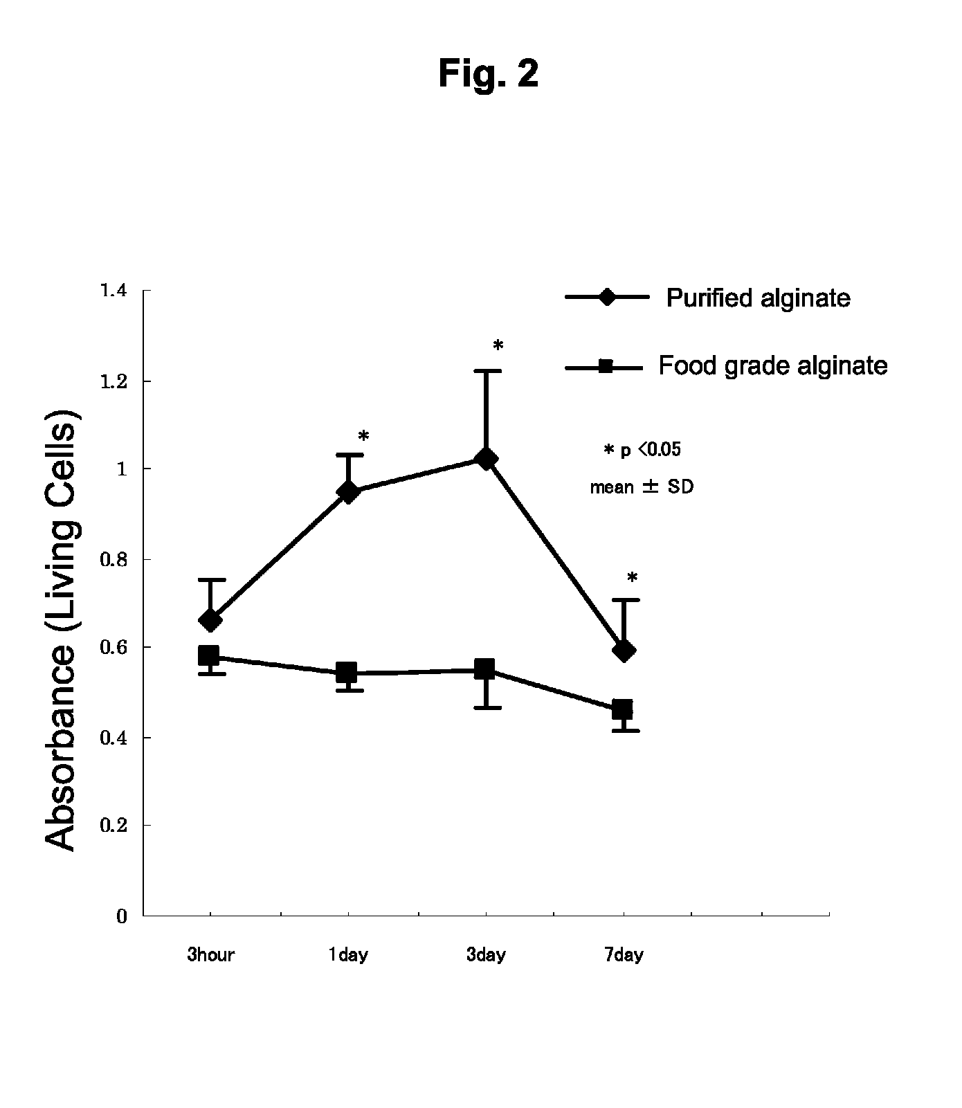
[0151]In the present example, two types of sodium alginate were used consisting of purified sodium alginate (Kimica Corp., Mochida International Ltd., Sea Matrix (sterilized), Serial No. B5Y01) and non-purified, food grade sodium alginate (also referred to as commercial grade sodium alginate, Wako Pure Chemical Industries, Ltd., Sodium Alginate 500, 199-09961). The purified sodium alginate was sterilized and freeze-dried. The food grade sodium alginate was sterilized by filtering with a filter having a pore diameter of 0.22 μm.

[0152]When endotoxin levels were measured using a commercially available LAL assay kit (Limulus Color KY Test Wako, Wako, Japan), the endotoxin level of the purified sodium alginate was 5.76 EU (endotoxin units) / g and that of the food grade sodium alginate was 75950 EU / g, thus indicating that the endotoxin level of the purified sodium alginate was far lower than that of the food grade sodium alginate. Namely, the purified...

example 2

Production of Transplant Cells

[0160]Bone marrow mesenchymal stromal cells (BMSC) were isolated and cultured to obtain transplant cells. BMSC include erythropoietic cells and the like in addition to bone marrow mesenchymal stem cells. 1 mL of bone marrow were harvested from the tibia of four-month-old Japanese white rabbits followed by washing twice with Ca—Mg-free PBS (Gibco BRL Lab.) and suspending in DMEM-High Glucose (DMED-HCA Sigma Chemical, St. Louis, Mo.). Blood clots were removed with a cell strainer having a pore diameter of 70 μm (Falcon Co., Ltd.). The cells were then incubated while humidifying at 37° C. and 5% CO2 in a 100 mm culture dish containing a culture medium consisting of DMEM-HG, 10% fetal bovine serum (FBS, Gibco, Life Technology, Grand Island, N.Y.) and 1% antibiotics (Penicillin-Streptomycin-Fungizone 100× concentrated, Cambrex Biosciences, Walkersville, Md.). The culture medium was replaced every three days and non-adherent cells were removed. After monolaye...

example 3

Production of Alginate Beads

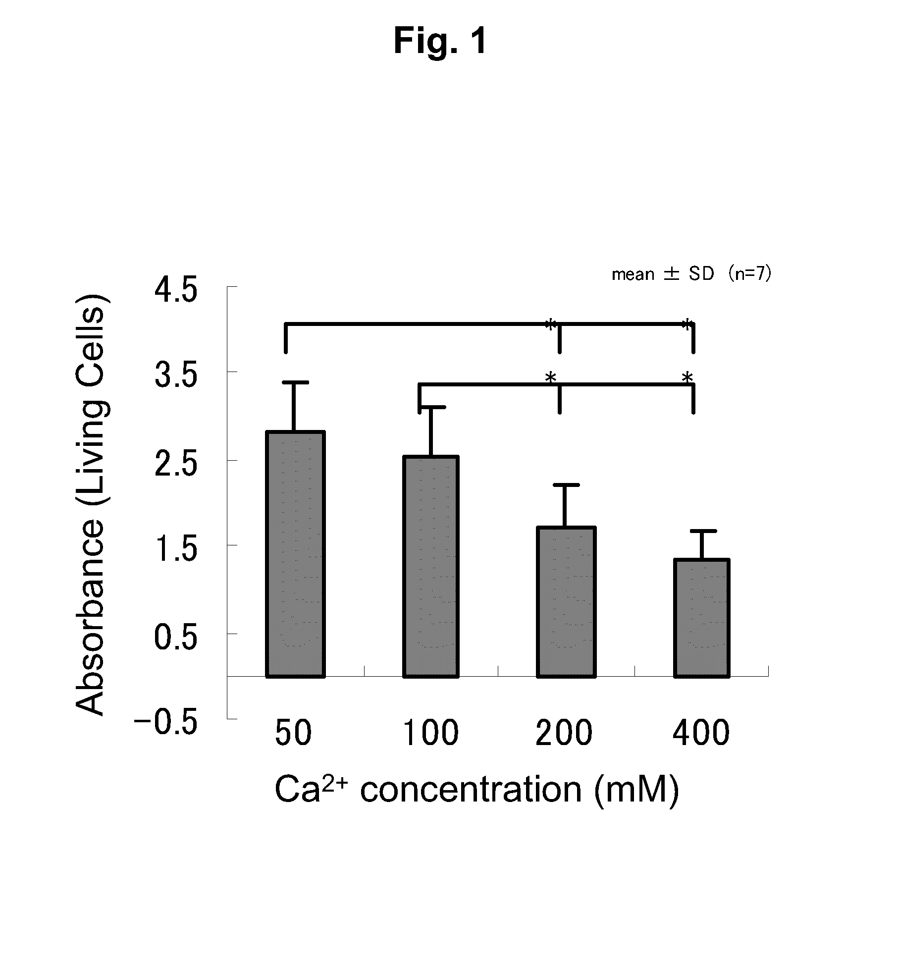
[0161]The cells obtained in Example 2 were suspended at 2.5×10⁷ cells / ml in a sodium alginate solution adjusted to a concentration of 2% w / v with filtration-sterilized Milli-Q water. The suspension was gelled by dropping into CaCl2 solution with a pipette, and after washing for two times the microcapsules that formed 10 minutes later with Ca—Mg-free PBS, the microcapsules were washed once with DMED-HG. The resulting beads contained 1×10⁶ cells per 40 μl per bead.

[0162]The cells were harvested from the alginate beads by washing three times with PBS and incubating at 37° and 5% CO2 in 50 mM EDTA (Gibco BRL Laboratories) followed by centrifuging for 5 minutes at 1500 g 10 minutes later to harvest the cells.


PUM

PropertyMeasurementUnit
viscosityaaaaaaaaaa
viscosityaaaaaaaaaa
viscosityaaaaaaaaaa

Abstract

The present invention provides a composition for regenerating cartilage or treating a cartilage disease containing a monovalent metal salt of alginic acid for which the endotoxin level thereof has been lowered to an extent that does not substantially induce inflammation or fever. As a result, it is possible to provide a composition for regenerating cartilage that improves cartilage regenerative action and ease of application to a cartilage injury lesion, and a composition for treating a cartilage disease, which has the effects of protecting cartilage from mechanical irritation, inhibiting degenerative changes in cartilage caused by wear and inflammation, repairing a cartilage injury lesion, and inhibiting inflammation and pain of joint tissue.

Description

CROSS-REFERENCE TO RELATED APPLICATIONS[0001]This application is a National Stage application of PCT / JP2008 / 052999, filed Feb. 21, 2008, which claims priority from Japanese applications JP 2007-041520, filed Feb. 21, 2007 and JP 2007-277005, filed Oct. 24, 2007.TECHNICAL FIELD[0002]The present invention relates to a composition for cartilage regeneration or cartilage disease treatment, including veterinary applications.BACKGROUND OF THE INVENTION[0003]For example, articular cartilage is hyaline cartridge that is composed of a small number of cells, collagenous extracellular matrix, abundant proteoglycans and water. In the case of bone, since vascular and neural networks are present and bone has the ability to self-repair, even if a fracture has occurred, the fracture is frequently completely repaired. However, articular cartilage lacks vascular and neural networks. Consequently, it has virtually no potential for self-repair, and in the case of the formation of large cartilage defect...

Claims


Application Information

Patent Timeline
Patent Type & Authority Patents(United States)
IPC IPC(8): A61K35/12A61F2/28A61F2/30A61K9/08A61K31/734A61K35/28A61K47/36A61L27/00A61L27/20A61L27/38A61P19/02A61P25/02A61P29/00C08B37/04C12N5/0775C12N15/09
CPCA61L27/20A61L27/3834A61L27/3852A61K35/28A61K31/734C08L5/04A61L2430/06A61P19/00A61P19/02A61P19/04A61P19/08A61P25/02A61P29/00
Inventor IWASAKI, NORIMASAMINAMI, AKIOKASAHARA, YASUHIKOIGARASHI, TATSUYAKAWAMURA, DAISUKEKASAHARA, FUMIYOSHIMIYAJIMA, CHIHIROOHZAWA, NOBUOIMAI, MARIKO
Owner MOCHIDA PHARM CO LTD
Why Eureka
Try Eureka