
Method and use of nano-scale devices for reduction of tissue injury in ischemic and reperfusion injury

a technology of nano-scale devices, which is applied in the direction of cardiovascular disorders, synthetic polymeric active ingredients, drug compositions, etc., can solve the problems of increased tissue stress, worsening damage, and inability and death, and achieve the effect of reducing ischemic and reperfusion injury

Inactive Publication Date: 2014-04-15
THE JOHN HOPKINS UNIV SCHOOL OF MEDICINE +1
View PDF17 Cites 0 Cited by
  • Summary
  • Abstract
  • Description
  • Claims
  • Application Information

AI Technical Summary

Benefits of technology

The patent describes methods for reducing damage caused by reduced blood flow (ischemic injury) in various tissues, including the brain, kidneys, and liver. The methods can also be used to protect tissue during surgeries and organ transplantations. The patent also describes a nanodevice that can be used to make a medicine that can be given to patients to reduce the damaging effects of ischemic reperfusion injury to tissue.

Problems solved by technology

Ischemic and reperfusion injury are major causes of disability and death in the United States.
Reperfusion may cause additional tissue stress, sometimes worsening damage.
Current strategies to protect tissues from the effects of ischemic and reperfusion damage are limited.
Such drugs reduce the causative factors involved in arterial blockage; however, they do not provide protection to a tissue affected by an ischemic event.
In addition, not all patients benefit from these treatments, due in part to factors including drug insensitivity, drug toxicities, and other risks (e.g. hemorrhage), and drug interactions.
These treatments may improve patient prognosis, however, tissue damage can occur from the procedure including the acute risk of vessel rupture and ischemic damage, and delayed risks include restenosis, or reocclusion of the occluded vessel leading to additional ischemic events.
In addition, there exists a need for nanodevices for the treatment of patients, including patients in which current pharmaceutical solutions are unacceptable.

Method used


Image

  • Method and use of nano-scale devices for reduction of tissue injury in ischemic and reperfusion injury
  • Method and use of nano-scale devices for reduction of tissue injury in ischemic and reperfusion injury
  • Method and use of nano-scale devices for reduction of tissue injury in ischemic and reperfusion injury

Examples


example 1

Nanodevices for Reducing Ischemic Injury

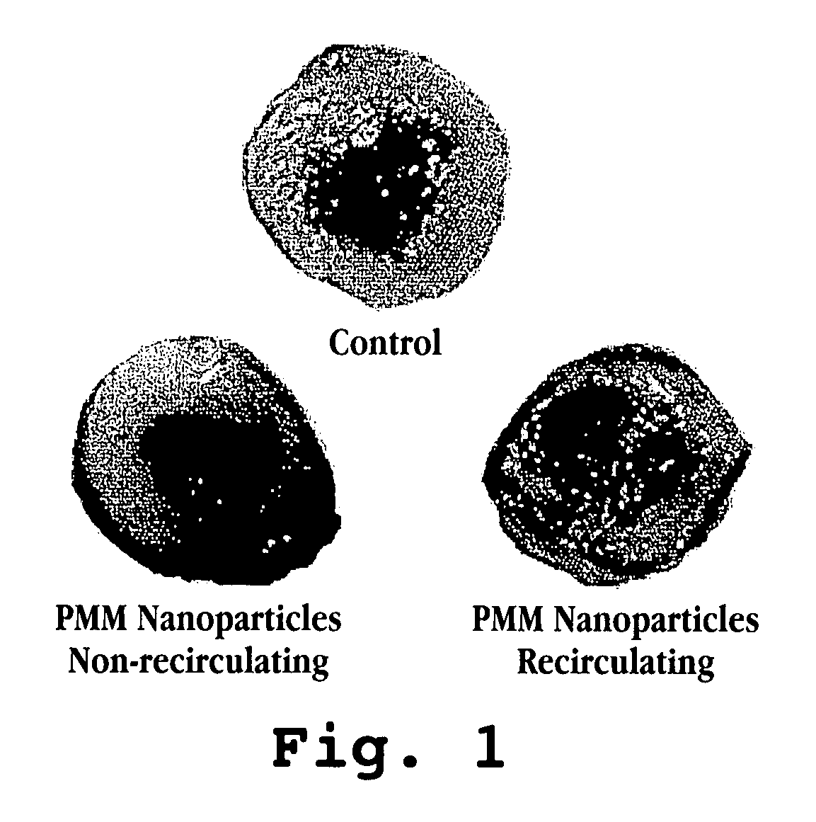
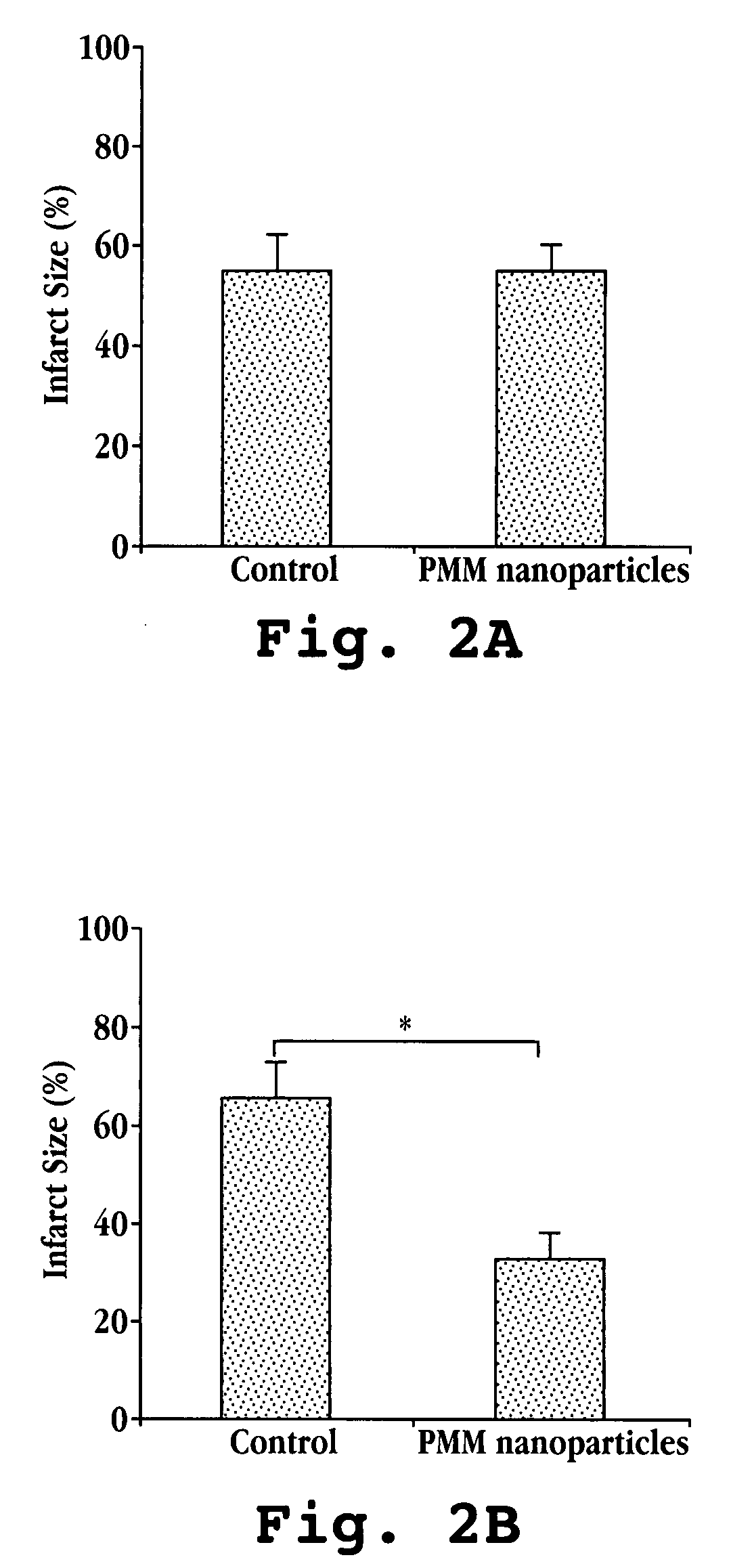
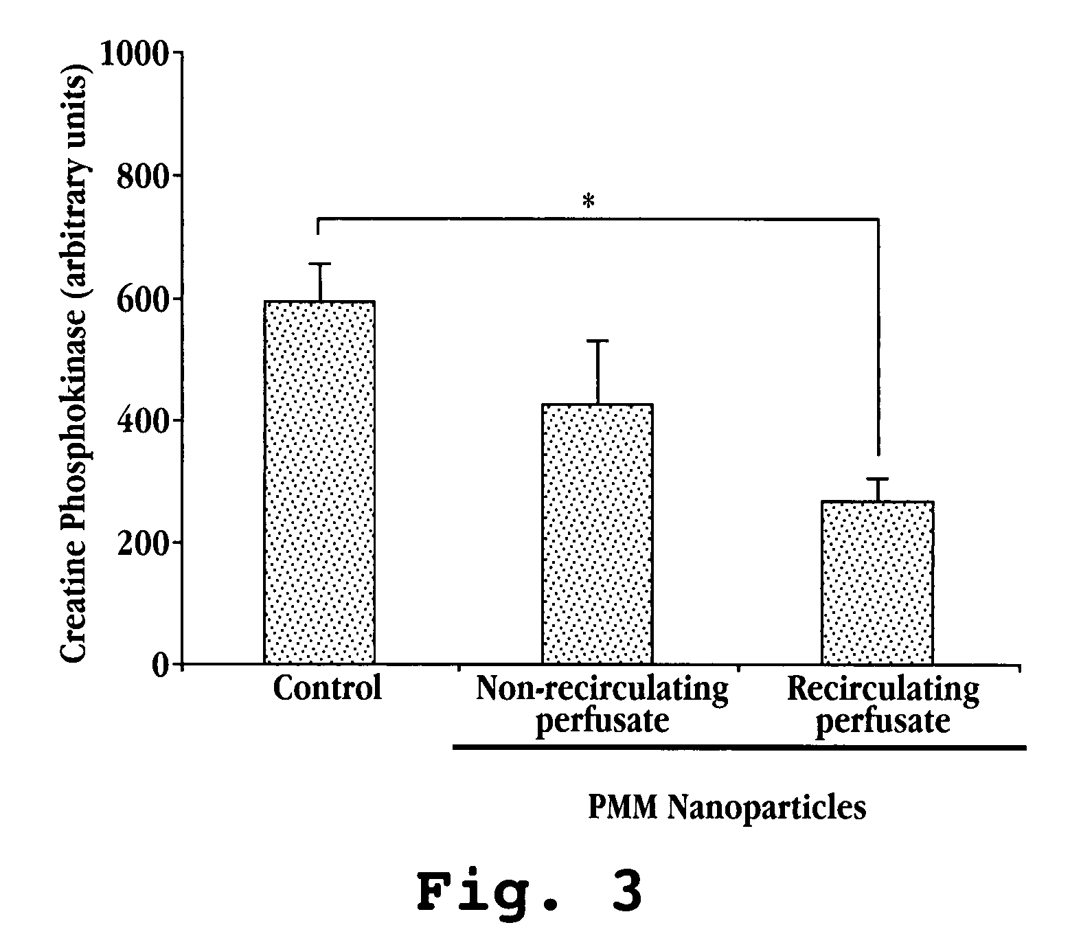
[0048]The present example shows that reduced tissue damage was achieved in a rat heart subject to ex vivo ischemia and reperfusion injury by treating the heart with nanoparticles.

[0049]A. Materials

[0050]Male Sprague-Dawley rats (280-380 g; n=4-5 per group) were used for ex vivo cardiac ischemia and reperfusion. PMM nanoparticles were synthesized via polymerization of poly(methylidene malonate 2.1.2 monomer as described previously (Breton, P. et al., Biomaterials, 19(1-3):271 (1998)). Polymer was delivered at 1×10⁶ particles / mL to the heart.

[0051]B. Methods

[0052]To mimic acute myocardial infarction, an ex vivo Langendorff cardiac ischemia / reperfusion model was used. Working hearts were extracted from deeply anesthetized animals and rapidly cannulized. Oxygenated, warmed Krebs buffer was perfused in a retrograde fashion through the hearts via the aorta to maintain tissue viability. To mimic ischemia, perfusion of the oxygenated buffer was stoppe...

example 2

Identification of Optimal Surface Characteristics of Nanodevices

[0058]Nanodevices having surface characteristic optimal for delivering and releasing therapeutic agents (i.e., drugs) are determined by testing a series of nanodevices, e.g., in the animal model described in Example 1.

[0059]In one experiment, the following nanodevices are tested.

[0060]

Nanodevices with differing surface characteristics will be evaluated forcardioprotective effectsSurface characteristicsSizeNameFluorophore (λ)Negative charge0.5 μmPMM 2.1.2580 / 605Positive charge0.2 μmFS amine580 / 605Positive charge1.0 μmFS amine505 / 515Carboxylate modified0.2 μmFS carboxylate580 / 605Carboxylate modified0.5 μmFS carboxylate580 / 605Carboxylate modified1.0 μmFS carboxylate580 / 605

[0061]Each nanodevice is tested as described, e.g., in Example 1, with results being determined by measuring the levels of cardiac enzymes released in the buffer effluent (a measure of oncosis) as well as by staining with triphenyl tetrazolium chloride (T...

example 3

Measuring Adenosine Concentration in Cardiac Tissue

[0064]Preliminary studies suggest that nanodevices protect allografts via an adenosine receptor-dependent mechanism. However, the short half-life of adenosine (<1 second in the blood) makes studying this mechanism problematic. An inhibitor of adenosine deaminase (the enzyme responsible for degradation of adenosine and used for snap freezing) is used to facilitate study the mechanism. Briefly, hearts are perfused with cardioprotective nanoparticles for 2 minutes. A cold 15 G needle is used to collect biopsy samples every 30 seconds. The biopsy samples are snap frozen in tubes containing extraction buffer (0.4 M perchloric acid) and 280 μM deaminase inhibitor (e.g., erythro-9(2-hydroxy-3-nonyl)adenine; EHNA. Following two freeze / thaw cycles, shaking, and centrifugation, the samples are loaded onto a reverse-phase HPLC column to separate adenosine from other nucleotide catabolites. Samples are normalized to an adenosine standard and th...


PUM

PropertyMeasurementUnit
diameteraaaaaaaaaa
timeaaaaaaaaaa
timeaaaaaaaaaa

Abstract

A method for protection of tissues subject to ischemic and / or reperfusion damage is provided. The method includes administering to the tissue a composition comprising nanodevices. The nanodevices can take the form of, for example, polymeric nanoparticles or lipidic nanoparticles. The nanodevices also find use in methods for reducing ischemic injury in tissue at risk of such injury, such as heart and brain tissue.

Description

CROSS REFERENCE TO RELATED APPLICATIONS[0001]The application claims the benefit of U.S. Provisional Application No. 60 / 796,790, filed May 1, 2006, incorporated herein by reference in its entirety.FEDERALLY-SPONSORED RESEARCH OR DEVELOPMENT[0002]This invention was made with Government support under contract HL052141 awarded by the National Institutes of Health. The Government has certain rights in this invention.TECHNICAL FIELD[0003]The subject matter described herein relates to methods for reducing ischemic and reperfusion-induced tissue injury by delivering nanodevices to achieve a therapeutic benefit under conditions of ischemia / reperfusion.BACKGROUND[0004]Ischemic and reperfusion injury are major causes of disability and death in the United States. Ischemia is caused by a blockage and cessation of blood flow to a region of tissue, and may occur in multiple tissues, including the heart in diseases such as myocardial infarction and the brain, such as in ischemic stroke. Reperfusion...

Claims


Application Information

Patent Timeline
Patent Type & Authority Patents(United States)
IPC IPC(8): A61K9/127A61K9/14
CPCA61K9/5138A61K31/765A61K9/14A61K31/78A61K45/06A61P9/10
Inventor BRIGHT, CORINNEBRIGHT, RACHELCHURCHILL, ERICLEONG, KAM W.MOCHLY-ROSEN, DARIA
Owner THE JOHN HOPKINS UNIV SCHOOL OF MEDICINE
Learn More