Drug eluting medical devices having porous layers
a technology of porous layers and medical devices, applied in the field of medical devices, can solve the problems of inferior cell adhesion and growth relative to textured surfaces, smooth surfaces by their nature do not allow cell in-growth, and achieve the effects of reducing or selective retention of therapeutic agents, improving cell adhesion, and improving cell proliferation
- Summary
- Abstract
- Description
- Claims
- Application Information
AI Technical Summary
Benefits of technology
Problems solved by technology
Method used
Image
Examples
example 1
[0081]Stainless steel stents, 24 mm, 4 mm diameter, Liberté™, available from Boston Scientific, are cleaned with an RF oxygen plasma for 5 minutes to remove surface contaminants. Polyethyleneimine (PEI), poly(allylamine hydrochloride) (PAH), and poly(sodium-4-styrene sulfonate) (PSS) are available from Aldrich. Each of these polyelectrolytes is provided in solutions having a concentration of 1 mM polyelectrolyte and 0.8 M NaCl. Negatively charged sulfate-stabilized polystyrene (PS) spheres of diameter 330 nm are available from MicroPartikel, Berlin, Germany. The PS particle suspension is 0.1% m / m in pure water. Using the different solutions, an initial four layers of PEI-PAH-PSS-PAH are coated on the stent surface by dipping for 10 minutes in the separate solutions and rinsing twice in pure water for 10 minutes. The fifth layer is made by dipping the stent into the solution having the PS spheres. After this, the stent is rinsed with water and dipped again in the PAH solution. This s...
example 2
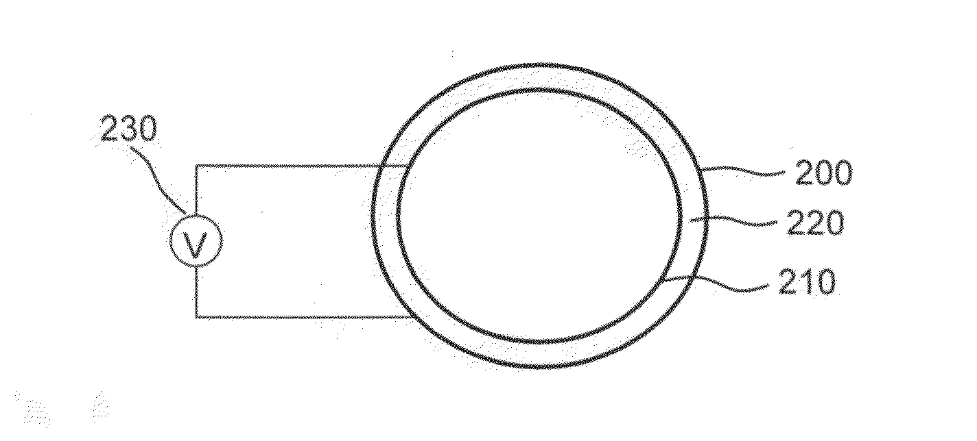
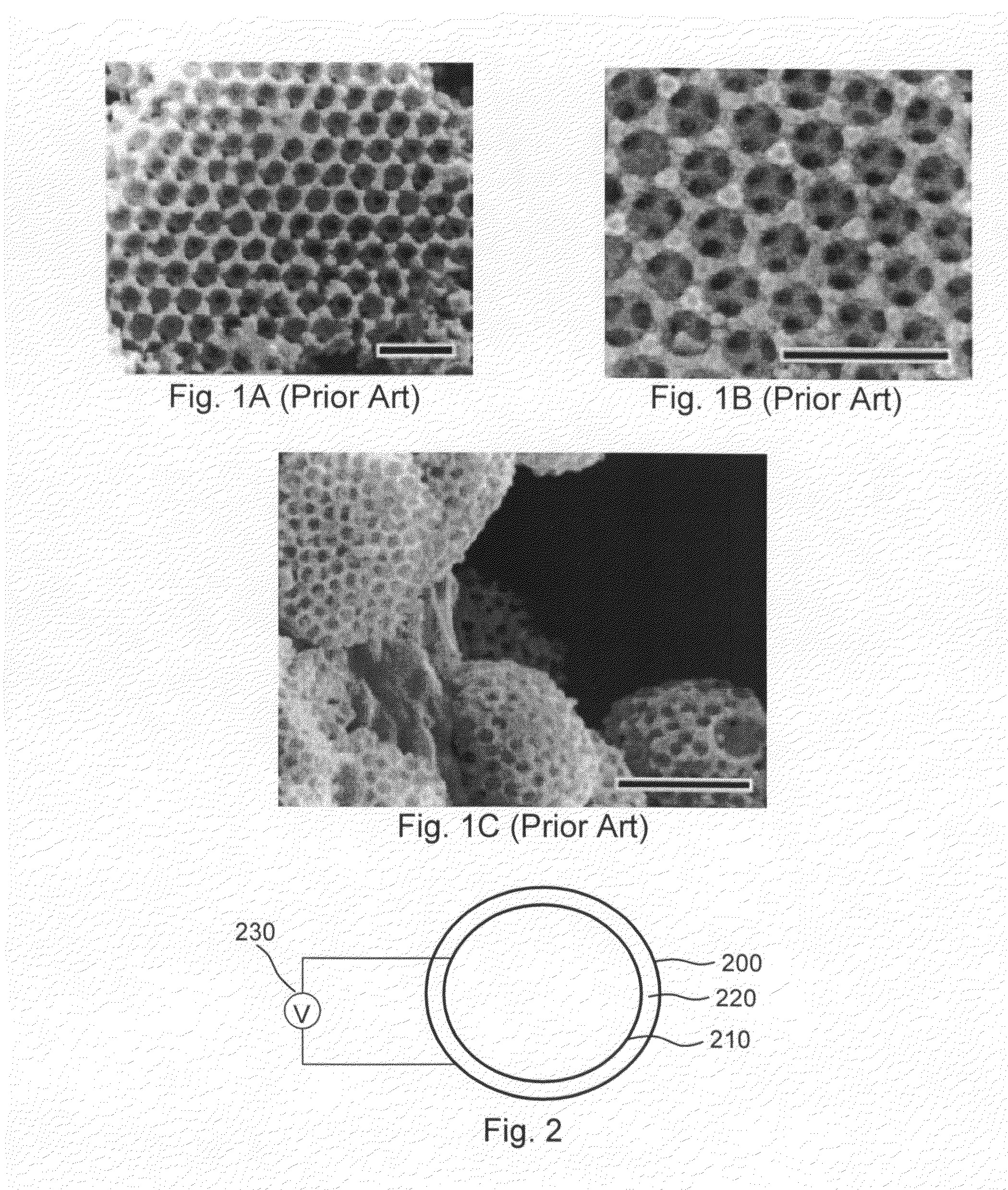
[0082]Stainless steel stents, 24 mm, 4 mm diameter, Liberté™, available from Boston Scientific, are cleaned with an RF oxygen plasma for 5 minutes to remove surface contaminants. Negatively charged sulfate-stabilized PS spheres of diameter 330 nm are available from MicroPartikel, Berlin, Germany. (Alternatively, negatively charged PS spheres of 975±10 nm diameter may be provided by Ikerlat Polymers as described in M. Yoldi et al., J. Mater. Sci., 41 (2006) 2965-2969.) Following procedures along the lines described in M. Yoldi et al., and using an electrochemical cell like that illustrated in FIG. 2, an aqueous colloidal dispersion of negatively charged polystyrene spheres in an aqueous-ethanolic medium is deposited on the interior surface of the stent, which corresponds to the positively charged electrode of the electrochemical cell, by applying a suitable DC voltage for a time sufficient to create a multilayer colloidal crystal (e.g., one having approximately 20 layers). Once the c...
example 3
[0083]Stents in accordance with Example 1 and Example 2 are subjected to sol-gel processing by immersing the stents for 12 hours in a solution of 2 wt % TEOS (tetra-ethoxy-silane), 88 wt % ethanol, 9 wt % water, and 1 wt % ammonium hydroxide. The stents are then calcinated at 540° C. for 8 hours to remove the organic components and cooled overnight in the oven to room temperature, yielding stents with a multi-layer structure of hollow silica spheres.
PUM
| Property | Measurement | Unit |
|---|---|---|
| diameter | aaaaa | aaaaa |
| diameter | aaaaa | aaaaa |
| sizes | aaaaa | aaaaa |
Abstract
Description
Claims
Application Information
 - R&D
- Intellectual Property
- Life Sciences
- Materials
- Tech Scout
- Unparalleled Data Quality
- Higher Quality Content
- 60% Fewer Hallucinations
Browse by: Latest US Patents, China's latest patents, Technical Efficacy Thesaurus, Application Domain, Technology Topic, Popular Technical Reports.
© 2025 PatSnap. All rights reserved.Legal|Privacy policy|Modern Slavery Act Transparency Statement|Sitemap|About US| Contact US: help@patsnap.com


