

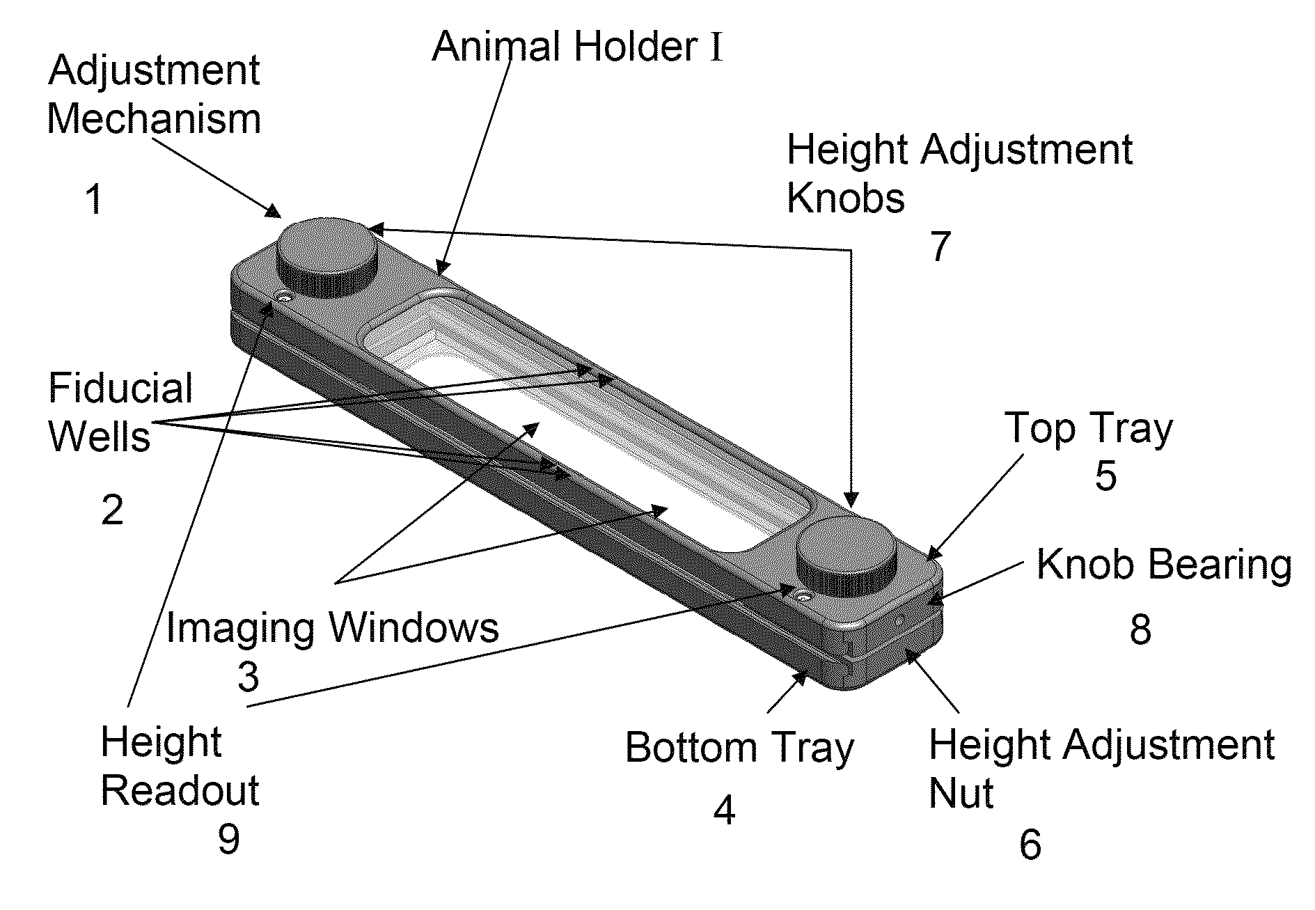
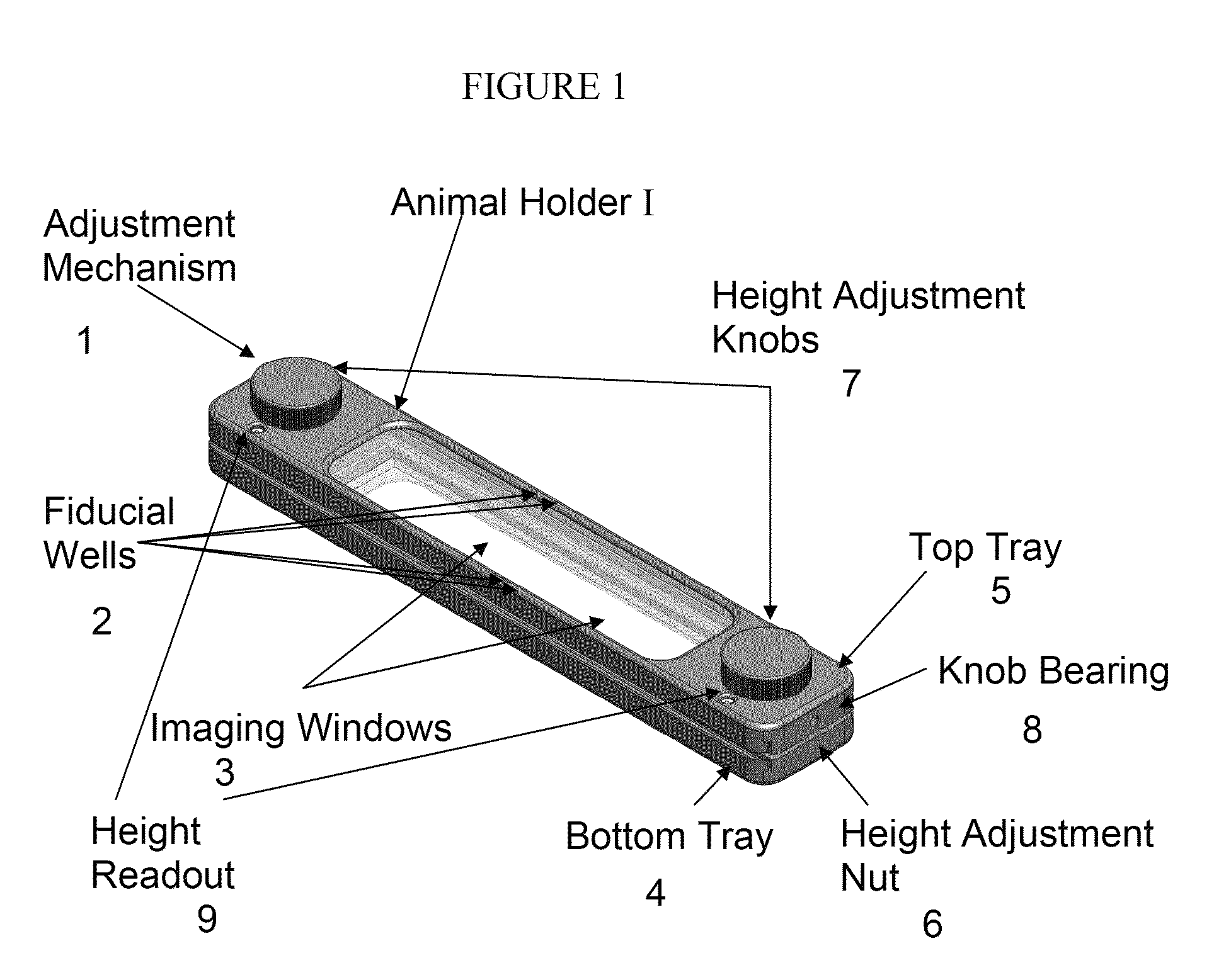
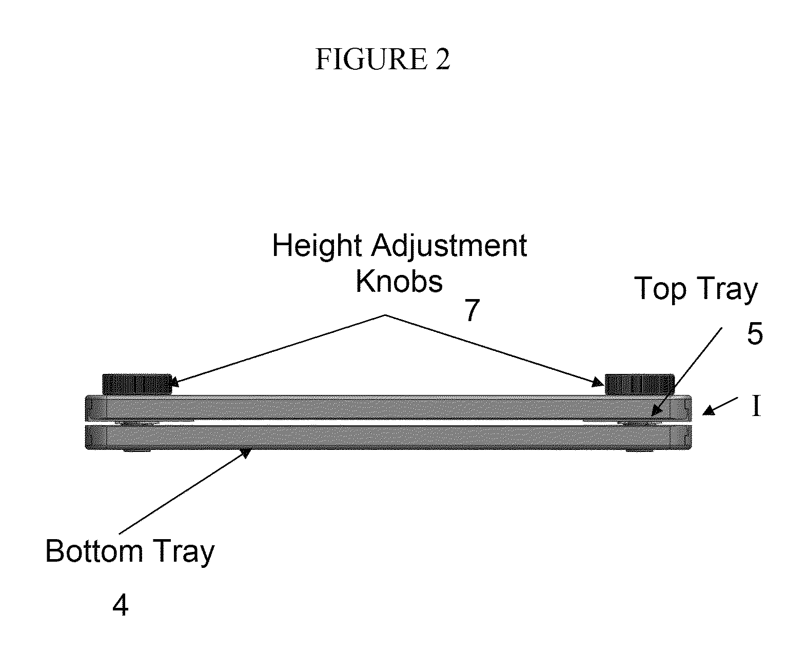
Animal holder for in vivo tomographic imaging with multiple modalities
a tomographic imaging and animal holder technology, applied in the field of in vivo tomographic imaging with multiple modalities, can solve the problems of insufficient information in the signal received by a single energy sensor (for example, at one angle) to generate either a two-dimensional or a three-dimensional representation of the internal structure of the object, and the existing x-ray cat system cannot provide functional (or, “molecular”) information about the subject or disease state at the cellular or mo
- Summary
- Abstract
- Description
- Claims
- Application Information
AI Technical Summary
Benefits of technology
Problems solved by technology
Method used
Image

Examples
example 1
Multi-modality Imaging using the Animal Holder
[0143]An example of multi-modality imaging is depicted in FIGS. 46-48. Alexa Fluor 680 (AF 680) dye (Invitrogen, Carlsbad, Calif.) was dissolved in water and injected into a plastic imaging phantom. The imaging phantom was surgically inserted subcutaneously into the thoracic cavity of an adult NU / NU mouse (Charles River Laboratories, Wilmington, Mass.). The mouse then was placed into an animal holder of the invention and secured for imaging before the entire cassette was placed inside an FMT2500 imaging system (VisEn Medical, Inc., Bedford, Mass.). Free dye dissolved in water (AF 680) was injected into the fiducial wells of the animal holder. An FMT imaging dataset was collected and subsequent reconstruction was performed using software included in the FMT2500. The animal holder containing the same mouse then was placed inside a 7 Tesla Bruker MR system (Bruker BioSpin, Billerica, Mass.) and an MR dataset was collected using Paravision 4...
PUM
 Abstract
Description
Claims
Application Information