Animal holder for in vivo tomographic imaging with multiple modalities

Active Publication Date: 2011-03-24
VISEN MEDICAL INC
View PDF18 Cites 38 Cited by
  • Summary
  • Abstract
  • Description
  • Claims
  • Application Information

AI Technical Summary

Benefits of technology

[0048]When an FMT modality is used, the light detected by the imaging system preferably includes excitation light from the light source that has been transmitted through the object and fluorescent light emitted from one or more fluorophores within the object. Data corresponding to the excitation light transmitted through the object, or intrinsic light, can be used to correct/calibrate captured fluorescent measurements, thereby providing more accurate tomographic images. The one or more fluorophores emit fluorescent light as a result of excitation by the excitation light. Background fluorescence can be accounted for by obtaining background measurements and processing data corresponding to the captured fluorescent light accordingly.

Problems solved by technology

A signal received by a single energy sensor (for example, at one angle) does not contain sufficient information to generate either a two-dimensional or a three-dimensional representation of internal structures of the object.
While X-ray CAT imaging is able to provide high resolution of certain anatomical structures, it is relatively limited in its ability to detect, distinguish, or quantify specific chemical or biological species in the subject.
Therefore, existing X-ray CAT systems cannot provide functional (or, “molecular”) information about a subject or disease state at the cellular or molecular level.
Specific molecular information with these modalities cannot often be obtained or is limited.
While optical tomography is well suited to providing molecular/functional information about a subject, the achievable resolution is not as high as with X-ray CAT o

Method used


Image

  • Animal holder for in vivo tomographic imaging with multiple modalities
  • Animal holder for in vivo tomographic imaging with multiple modalities
  • Animal holder for in vivo tomographic imaging with multiple modalities

Examples


example 1

Multi-Modality Imaging using the Animal Holder

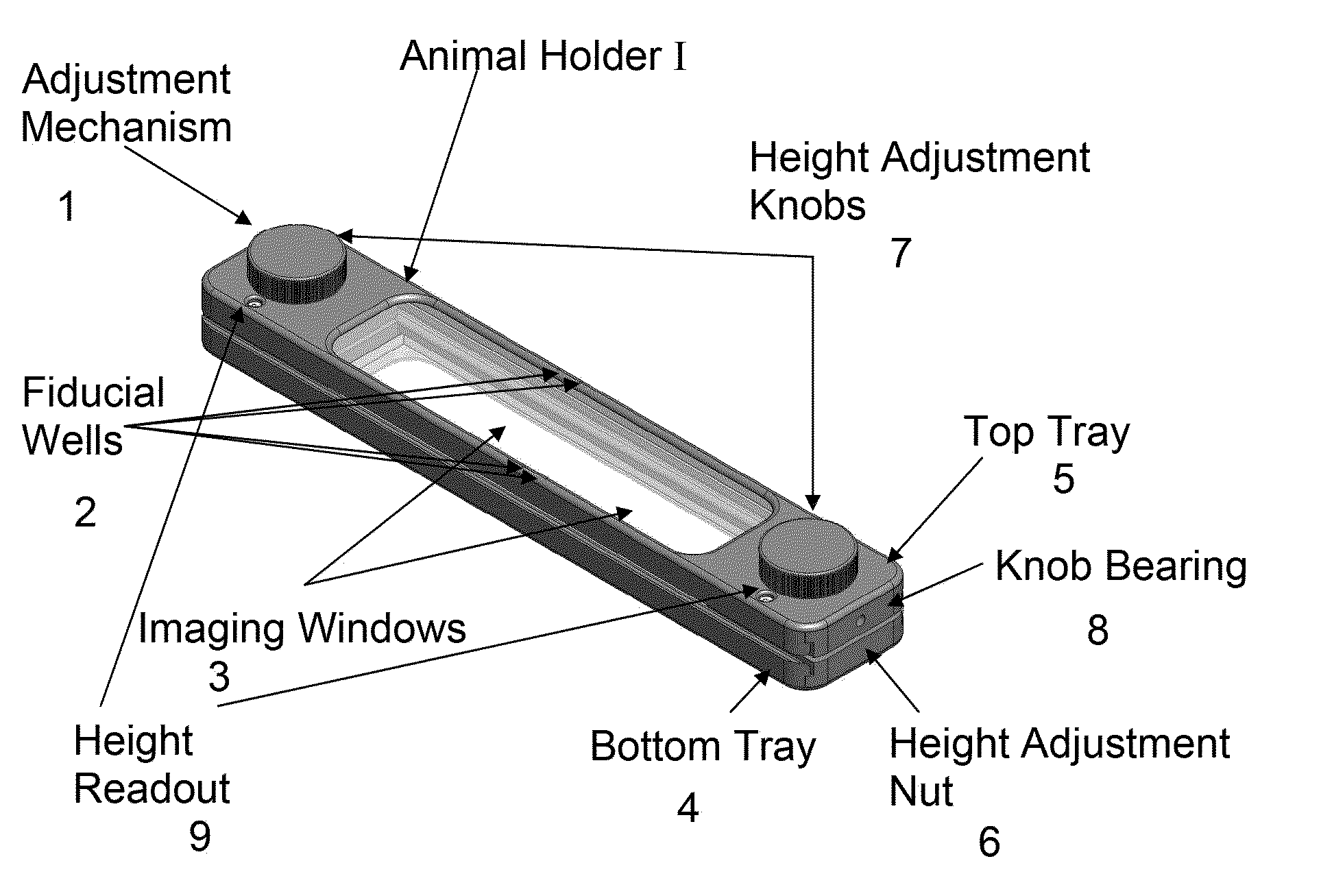
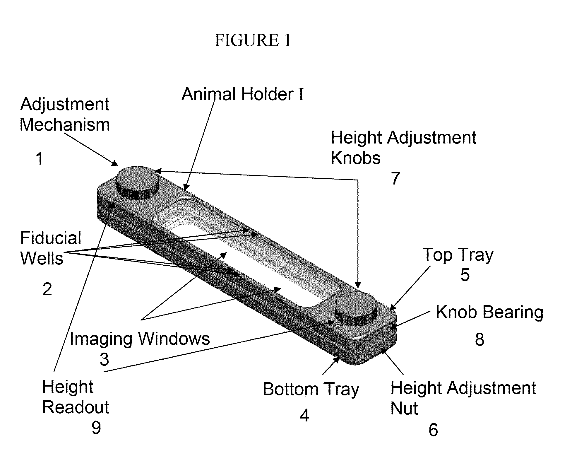
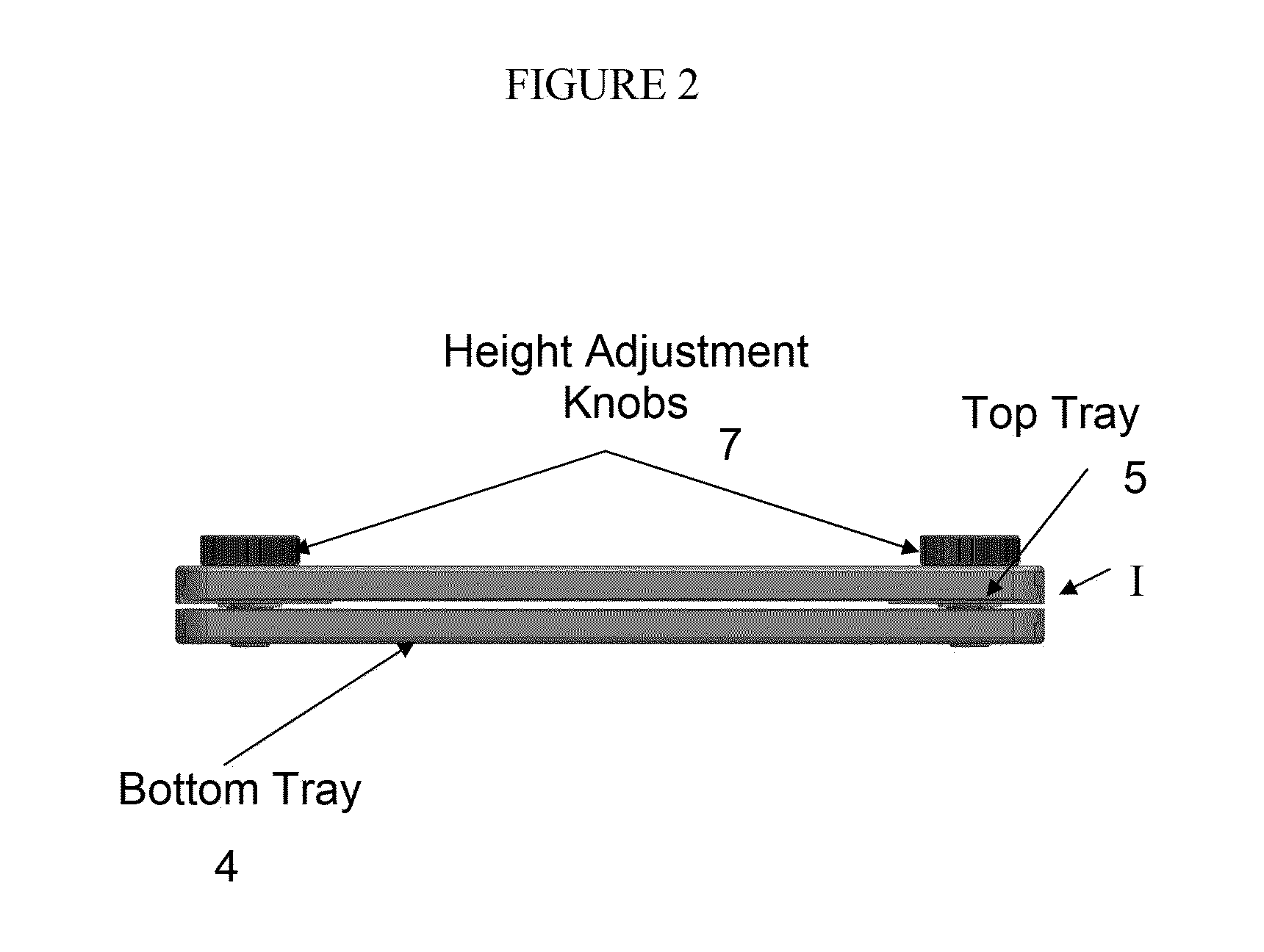
[0143]An example of multi-modality imaging is depicted in FIGS. 46-48. Alexa Fluor 680 (AF 680) dye (Invitrogen, Carlsbad, Calif.) was dissolved in water and injected into a plastic imaging phantom. The imaging phantom was surgically inserted subcutaneously into the thoracic cavity of an adult NU / NU mouse (Charles River Laboratories, Wilmington, Mass.). The mouse then was placed into an animal holder of the invention and secured for imaging before the entire cassette was placed inside an FMT2500 imaging system (VisEn Medical, Inc., Bedford, Mass.). Free dye dissolved in water (AF 680) was injected into the fiducial wells of the animal holder. An FMT imaging dataset was collected and subsequent reconstruction was performed using software included in the FMT2500. The animal holder containing the same mouse then was placed inside a 7 Tesla Bruker MR system (Bruker BioSpin, Billerica, Mass.) and an MR dataset was collected using Paravision 4...


PUM


Abstract

The invention facilitates transport of an immobilized, anesthetized small animal across multiple single-modality or multiple-modality imaging workstations at the same or different physical locations without loss of subject positional information. The animal holder is compatible with preclinical animal imaging stations such as micro-CT, micro-MR, micro-PET, micro-SPECT, and FMT. The animal holder is configured to be accommodated by (for example, fit within) individual imaging chambers of such instruments and is fabricated from materials that are compliant with all of the imaging modalities used. In certain embodiments, an integrated set of fiducial marker wells accommodates the dispensing of markers that are picked up by several modalities simultaneously in multiple planes. The fiducial markers then are aligned in standard image processing or image analysis software with simple image translation and rotation operations, without the need for more advanced scaling, distortion or other operations. The animal holder optionally also includes a system for providing the animal with inhalation anesthesia, for example, isoflurane, and is designed to fit into identical receptacles inside or outside the imaging workstation(s) that provide heating to minimize or prevent animal hypothermia.

Description

CROSS-REFERENCE TO RELATED APPLICATIONS[0001]This application claims the benefit of and priority to co-pending U.S. Provisional Patent Application No. 61 / 039,377, filed Mar. 25, 2008, the entire contents of which are incorporated by reference herein.GOVERNMENT RIGHTS[0002]The invention was supported, in whole or in part, by grant 1 R44 ES012699-01 of the National Institute of Environmental Health Sciences. The Government has certain rights in the invention.FIELD OF THE INVENTION[0003]The invention relates generally to animal restraining systems for in vivo imaging across multiple tomographic modalities and / or imaging systems. More particularly, in certain embodiments, the invention relates to an animal holder compatible with optical imaging systems in conjunction with magnetic resonance, computed tomography, positron emission tomography, and / or other tomographic imaging systems, to enhance and simplify the registration of reconstructed datasets acquired on different systems.BACKGROU...

Claims


Application Information

Patent Timeline
IPC IPC(8): A61B6/00A01K15/04
CPCA61B5/7285A61B6/0421A61B6/045A61B6/583A61B8/543A61B2503/40A61B6/508A61B6/485
Inventor YARED, WAEL I.WILSON, ANDREW K.
Owner VISEN MEDICAL INC
Why Eureka
Try Eureka