

Microvascular stamp for patterning of functional neovessels
- Summary
- Abstract
- Description
- Claims
- Application Information
AI Technical Summary
Benefits of technology
Problems solved by technology
Method used
Image


Examples
example 1
Hydrogel Preparation
[0079]Gel-forming polymers, poly(ethylene glycol) diacrylate (PEGDA) and methacrylic alginate (MA), were synthesized as described in FIG. 6 and our previous works16. Briefly, for the synthesis of MA, 2-Aminoethylmethacrylate was conjugated to the carboxylate group of alginate via EDC chemistry. See FIG. 6a. The alginate used in this experiment (molecular weight (Mw)˜50,000 g / mol) was obtained by irradiating alginate rich in gluronic acid residues, (LF20 / 40, FMC Technologies, Mw˜250,000 g / mol) with a dose of 2 Mrad for 4 hours from a 60Co source. The irradiated alginate was dissolved in the 0.1M MES ((2-(N-morpholino) ethanesulfonic acid) buffer (pH 6.4, Sigma-Aldrich) at the concentration of 1.0% (w / v). Then, 1-hydroxybenzotriazole (HOBt, Fluka), 1-ethyl-3-(3-dimethylaminopropyl) carbodiimide (EDC, Thermo Scientific) and 2-aminoethyl methacrylate (AEMA, Sigma Aldrich) were dissolved in the alginate solution and stirred for 18 hours. Both the molar ratio of HOBt t...
example 2
Cell Encapsulation
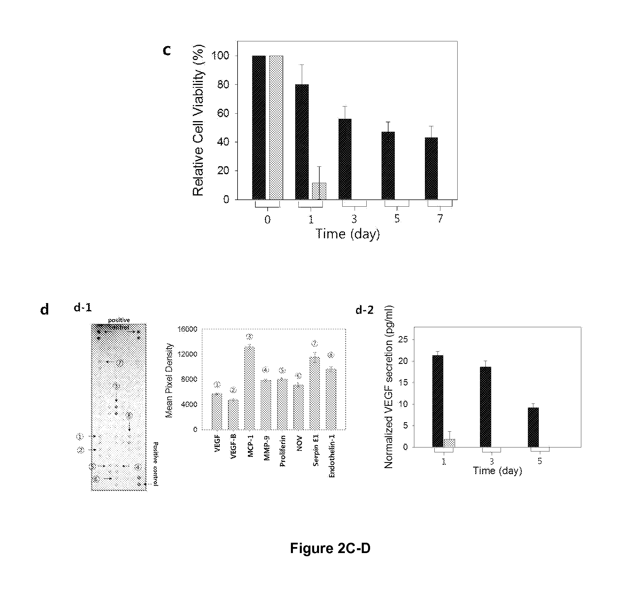
[0094]NIH / 3T3 cells (ATCC) were expanded and passaged at 37° C. with 5% CO2 in Dulbecco's modified Eagle's medium (DMEM, ATCC) supplemented by 10% fetal bovine serum (FBS, ATCC), and 1% penicillin / streptomycin (ATCC). All the cells before the passage number of 10 were used in this study. Prior to encapsulation in hydrogels, cells were mixed with the pre-gelled polymer solution. The cell density was kept constant at 2.0×10⁶ cells / ml. The mixture of cell and pre-gelled solution was exposed to the laser of SLA to activate hydrogel formation. (FIG. 13). NIH / 3T3 cells cultured for seven days while being encapsulated within hydrogels. A uniform distribution of cells was achieved throughout the hydrogel by the bottoms-up approach of SLA. FIG. 13a. The viability of encapsulated cell was evaluated by adding 0.1 mL of a growth medium and 0.01 mL of MTT (3-(4,5-dimethylthiazol-2-yl)-2,5-diphenyltetrazolium bromide) reagent (ATCC) into a well of a 96-well plate which contained...
example 3
Microchannels
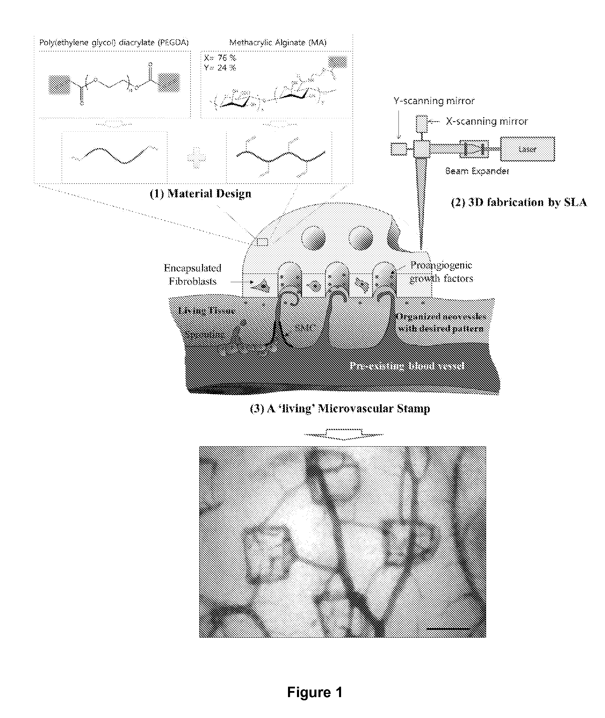
[0099]Next, microchannels with controlled spacing were introduced into the PEGDA-MA hydrogels with the goal of driving neovessel growth along its circular pattern (FIG. 12). Neovessel growth direction can be controlled by increasing the flux of the cell-secreted proangiogenic factors through the microchannel wall, so that the neovessels localize within the microchannel lumen (FIG. 3a). According to Fick's law of diffusion (Eq. (1) above), the ratio between the flux of growth factors through the microchannel wall of the stamp (Jm) and the flux through the bottom of the stamp with the same cross-sectional area as the microchannel (Jf) is equal to the ratio between the surface area of microchannel wall (Swall) and the cross-sectional area of the microchannel (Sbottom). Hence, for a hydrogel with thickness of 200 μm, it could be predicted that the diameter of the microchannels should be less than 800 μm in order to make Jm larger than Jf.
[0100]Based on this prediction, micr...
PUM
| Property | Measurement | Unit |
|---|---|---|
| Thickness | aaaaa | aaaaa |
| Thickness | aaaaa | aaaaa |
| Diameter | aaaaa | aaaaa |
Abstract
Description
Claims
Application Information