
Noninvasive detection of meconium in amniotic fluid

a technology of amniotic fluid and meconium, which is applied in the field of noninvasive detection of meconium in amniotic fluid, can solve the problems of obstructing airways, meconium aspiration syndrome (mas), and 1000 children's deaths, and achieve the effect of saving critical time and high amniotic fluid sugar level

Active Publication Date: 2015-09-29
MOR GIDEON
View PDF24 Cites 0 Cited by
  • Summary
  • Abstract
  • Description
  • Claims
  • Application Information

AI Technical Summary

Benefits of technology

Enables real-time detection of meconium secretion, facilitating early identification of fetal distress and reducing medical mismanagement by providing a simple, accurate method for laypeople to detect meconium-stained amniotic fluid, both before and after rupture of membranes, and until birth.

Problems solved by technology

Fetal stool, meconium, is toxic in the lungs, it can obstruct airways, and also does harm by other mechanisms, resulting in meconium aspiration syndrome (MAS).
About 13% of all live births are complicated by meconium-stained amniotic fluid (MSAF), and about 30,000 develop MAS annually in the United States, leading to the death of 1000 children.
MSAF is associated with an increased risk for perinatal complications during labor and delivery (e.g., low Apgar score, higher risk for cesarean section, and higher admission rate to neonatal intensive care unit.
Amnioscopy is a less invasive technique, enabling direct observation of the forebag of an amniotic sac to look for meconium staining; it may help in detecting heavy staining by meconium, but milder cases remain undetected, and the method may require an undesirable degree of cervical dilation.
All approaches described above must be employed by professional personnel and, therefore, limit their use to medical facilities.
As a matter of fact, high quality fetal wellbeing monitoring is still restricted to skilled personnel (e.g., ultrasonographic fetal heart rate tracing, biophysical profiling, and amniotic fluid indexing).
During this process the UC loop may be compressed and result in imminent fetal asphyxia.
Failure to recognize and immediately treat these complications may result in fetal death.
To date, there is no simple accurate method or device that enables home detection of MSAF by lay people.
However, in a significant number of events MSAF may be bloody or may be lightly stained and therefore not detectable by the naked eye.
To date, there is no simple method or device enabling such a thing.

Method used


Image

  • Noninvasive detection of meconium in amniotic fluid
  • Noninvasive detection of meconium in amniotic fluid
  • Noninvasive detection of meconium in amniotic fluid

Examples


example 1

[0060]After obtaining informed consent, amniotic fluid specimens were collected from women in labor following rupture of membranes. Urine specimens were collected through a urethral catheter from time to time during and after labor.

[0061]Phase I of this experiment was performed in order to determine whether detectable levels of CEA and / or AFP may be found and whether these levels may have diagnostic value. Phase I was performed as follows: Amniotic fluid specimens were examined by medical personnel and were classified as clear amniotic fluid or meconium stained amniotic fluid (MSAF). Samples of clear amniotic fluid and samples of MSAF were then sent for quantitative measurement of CEA and AFP concentrations by ABBOTT AxSYM (a Microparticle Enzyme Immunoassay, ABBOTT Laboratories, Ill., USA).

[0062]Table 2 shows results of the quantitative measurements of CEA and AFP.

[0063]

TABLE 2CEAAFPSAMPLE TYPEng / mLng / mL1Clear AF91140.752Clear AF145.6196.693Clear AF118.3291.124Clear AF209.3150.515C...

example 2

Amniotic Fluid and MSAF Detection

[0097]A commercial kit for detecting bile acid was purchased from Diazyme Laboratories (Poway, Calif., USA). Deoxycholic acid (DOC, a bile salt) was purchased from Sigma-Aldrich (St. Louis, Mo., USA). Accurate implementation of the procedure described by the Diazyme kit manual showed that the presence of DOC can be easily detected by the naked eye. DOC was applied to the solution containing the reconstituted kit reagents at room temperature. The original solution color is yellow. The presence of DOC initiated a two-stage reaction that led to a color change to dark red / brown after ten minutes. The presence of low DOC concentrations could be easily detected by the naked eye. Controls containing distilled water did not lead to any color change. Solutions maintained a stable color for approximately one hour. Meconium bile acids can be detected by the color change. A pH sensitive pad for the detection of amniotic fluid (Common-Sense Inc., Caesarea, Israel...

example 3

A Platform for Identifying Etiologies of Fetal Distress after ROM Occurs

[0098]High amniotic glucose level can be a result of uncontrolled gestational diabetes, which is a possible etiology for fetal distress. A colorimetric assay for glucose can be done by coupling the activities of the two enzymes: glucose oxidase and glucose peroxidase. In the first step, glucose oxidase catalyzes the reaction: glucose+oxygengluconolactone+hydrogen peroxide. In the second step, the hydrogen peroxide is used by the peroxidase to convert a chemical substrate (called a chromogen) from an uncolored form to a colored one. This reaction is used in commercial urinalysis kits. For example: the enzymes and the chromogen can be attached to the stick. When glucose is present, the reactions described above take place, and the stick changes color. In cases of early preterm premature rapture of membranes (PPROM; <32nd gestational week), the chances of placental abruption are as high as 10-15%. Benign abruption...


PUM

PropertyMeasurementUnit
air humidityaaaaaaaaaa
acid-baseaaaaaaaaaa
thresholdaaaaaaaaaa

Abstract

Meconium stained amniotic fluid can indicate intrauterine fetal distress. The invention provides a system and method for detecting meconium in released amniotic fluid in pregnant women, including a collection body such as a hygienic pad, having a meconium detector. This invention is designed to be employed mainly by a lay person at home; it also may provide possible etiologies for fetal distress.

Description

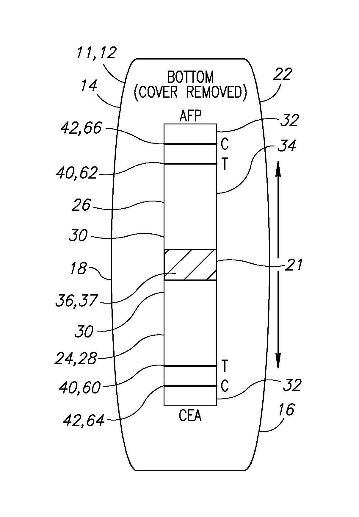
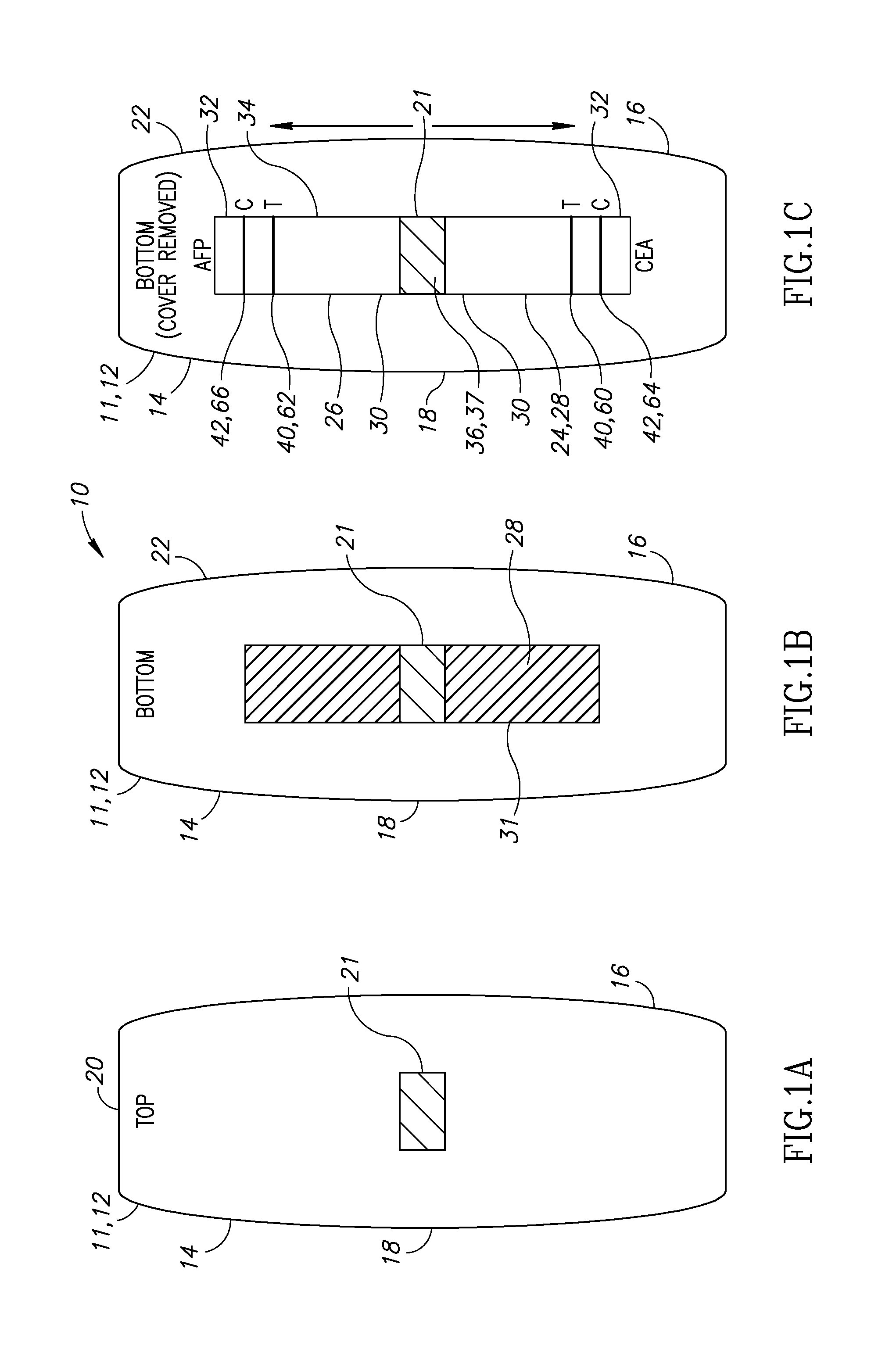
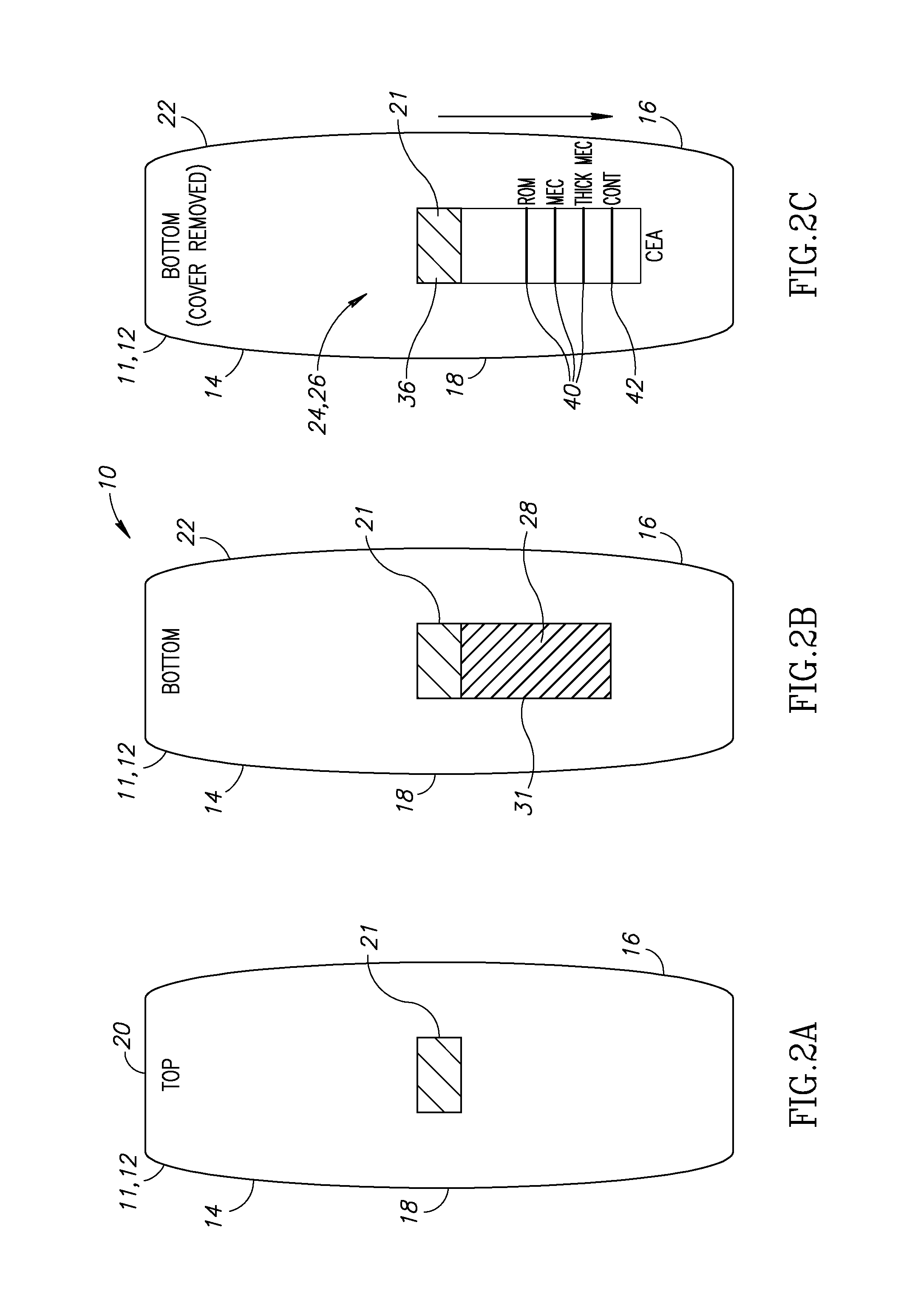
CROSS-REFERENCE TO RELATED APPLICATIONS[0001]This application is a continuation-in-part of International Application No. PCT / IL2012 / 050243, filed on Jul. 11, 2012, which claims the benefit of priority of Israel Patent Application No. 214,096, filed on Jul. 14, 2011, and which are incorporated herein by reference in their entirety.FIELD OF THE INVENTION[0002]The present invention relates to a system and method for detecting meconium in released amniotic fluid in pregnant women.BACKGROUND OF THE INVENTION[0003]The colon of a fetus during gestation fills with meconium, the fetal feces comprising bile and its metabolites, gastrointestinal and pancreatic secretions, mucus, cellular debris, swallowed vernix caseosa, and blood. In a significant minority of births, the fetus ceases to be continent and excretes meconium into the amniotic fluid. It is important to be aware of such an occurrence for the future mother and for the obstetrics personnel.[0004]The fetus may pass stool to the amniot...

Claims


Application Information

Patent Timeline
Patent Type & Authority Patents(United States)
IPC IPC(8): A61M1/00A61B5/00A61F13/505A61F13/42A61B10/00
CPCA61F13/42A61B5/4294A61B5/4362A61B10/0048A61F13/505A61B2503/02G01N33/689A61F13/20A61B5/14546A61B5/14503A61B5/14507A61B2562/0295G01N2333/90638G01N2333/70596G01N2400/00
Inventor MOR, GIDEON
Owner MOR GIDEON
Learn More