Scalable bio-element analysis

a bioelement and element technology, applied in the field of scalable bioelement analysis, can solve the problems of difficult to establish the precise origin of a positive signal, loss of potential binding candidates, and low screening efficiency, and achieve the goal of facilitating high throughput screening techniques to selectively isolate and purify identified clones for rapid application to disease diagnostics and therapeutics.

Active Publication Date: 2017-05-23
THE BOARD OF TRUSTEES OF THE LELAND STANFORD JUNIOR UNIV
View PDF20 Cites 31 Cited by
  • Summary
  • Abstract
  • Description
  • Claims
  • Application Information

AI Technical Summary

Benefits of technology

[0028]The invention provides several advantages over other methods of screening populations of biological elements. First, it allows simple screening of millions, billions or more, of biological interactions in parallel. The invention also allows for the display and independent recovery of analytes, such as target cells. In addition, it provides concentration versus affinity information for billions of clones in parallel (e.g., feedback on production efficiency can be provided for each expressed gene).

Problems solved by technology

Such systems have a number of disadvantages, including the need to enrich for desired clones via repeat selection steps (including, for example “panning”) that inherently result in the loss of potential binding candidates.
It is also difficult to establish the precise origin of a positive signal using conventional technologies since they obtain mixed signals from heterogeneous populations that cannot be deconvoluted.
While spatial addressing can, for example, identify specific cellular clones to generate master stocks, these approaches do not facilitate high throughput screening techniques to selectively isolate and purify the identified clones for rapid application to disease diagnostics and therapeutics.
Another disadvantage of the present screening assays is that they are usually limited to a cell number between approximately 50 and 100 thousand.
For performing cell-based screening, one of the particular challenges is the isolation of small populations of cells in a manner that allows for subsequent screening procedures.
Traditional devices and methods of isolating cells do not adequately provide for the isolation of small populations of cells without performing steps that potentially modify cellular function or activity.
Current screening methods have numerous technical challenges, including: the size of protein displayed has to be small, the multiplicity of infection (MOI) needs to be high to avoid loss of diversity, the dependency on the activity of the phage, multiple panning rounds are needed (taking up to 1 week or more), high non-specific binding due to phage, antibodies may not function well in soluble form (truncated clones are often expressed), and / or avidity effects can hinder selection of high affinity clones.

Method used


Image

  • Scalable bio-element analysis
  • Scalable bio-element analysis
  • Scalable bio-element analysis

Examples


example 1

Magnetic Bead Signal Blocking

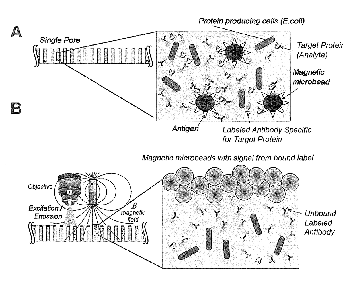
[0178]This example demonstrates the ability of the magnetic beads to inhibit the high background signal from labels in solution that are not bound to the particles.

[0179]A clean and dry 20 μm pore diameter array plate (J5022-19, Hamamatsu Photonics K. K.) was loaded with 10 μM Alexa 488 fluorochrome (Sigma Aldrich) as a label and 2.8 μm magnetic particles such that each pore receives both labels and magnetic particles. The fluorochrome label alone does not bind or associate with the magnetic bead particles.

[0180]The pores of the array are not exposed to any motive force prior to detection, so both the magnetic particles and the labels both remain in solution. Following incubation, this array comprising the magnetic particles and labels is subjected to EM radiation and the fluorescent emission from the label is imaged using a microscope. As expected, as shown in FIG. 5A, all of the pores show significant signal, for the unbound labels in solution have ful...

example 2

Assay Preparation

[0182]This example provides a representative assay system for detecting the presence or absence of an analyte in an array. The final screening design will, of course, depend on many factors specific to the experiment and its objectives.

[0183]The E. coli strain Imp4213, which contains a plasmid expressing GFP, was used in this assay. Approximately 1.0 to 10 million GFP-expressing bacterial cells were added into 5 mL of Luria broth (LB) media with 25 μg / ml kanamycin (KAN) and expanded on a shaker (200 RPM) at 37° C. for approximately 3 hours until the bacteria entered the exponential growth phase, and then were prepared for plating.

[0184]The particles used in this example are magnetic beads. Specifically, for these examples, the particles used were 2.8 μm diameter magnetic beads (Dynabeads) coupled to a either streptavidin (Part No. 112-05D, Invitrogen, Carlsbad, Calif.) or oligo(dt)25 ligands (Part No. 610-05, Invitrogen, Carlsbad, Calif.). The magnetic beads were in...

example 3

Single Cell Specific Detection

[0194]The following example demonstrates the specificity of the disclosed methods for detecting analytes at the single cell level.

[0195]In this embodiment, the assay system is set up according to the steps described in Example 2, except that before the cells are plated, the bacterial cells were diluted to 300 cells per microliter, which was calculated to result in approximately 0.1 cell per pore on the array plate with pores having a 20 micrometer diameter. When added to the array, this dilution will yield up to a single bacterial cell in most pores.

[0196]The anti-GFP streptavidin beads and anti-GFP label were added to the first array as described. The corresponding signal seen when EM radiation is exposed to the array following magnetic bead accumulation is shown in FIG. 7A. Many cells are signal-producing, which represents fluorescent signal from streptavidin magnetic beads that are bound to biotinylated anti-GFP antibody via streptavidin-biotin linka...


PUM

PropertyMeasurementUnit
diameteraaaaaaaaaa
diameteraaaaaaaaaa
diameteraaaaaaaaaa

Abstract

A method is provided for detecting one or more analytes in a sample. The method relies, in part, on the ability of functionalized particles added to the sample to partially or completely inhibit the transmission of electromagnetic radiation into and out of the sample through a detection surface in a reaction vessel containing the sample. In a microarray format, the invention can be used to screen millions, billions or more biological elements, such as an organism, cell, protein, nucleic acid, lipid, saccharide, metabolite, or small molecules. Methods, apparatuses and kits are described.

Description

CROSS REFERENCE TO RELATED APPLICATION[0001]This application claims the benefit of U.S. Provisional Patent Application Ser. No. 61 / 667,930 filed Jul. 3, 2012, which is incorporated by reference herein in its entirety.BACKGROUND[0002]Technical Field[0003]The invention relates to the detection of one or more analytes in a sample or a series of samples. More particularly, the invention relates to a method for determining one or more biological elements, e.g., biological cells, in a population of biological elements.[0004]Related Art[0005]Using conventional technologies, biological libraries may be screened for components containing, for example, cells, antibodies, proteins, peptides and, nucleic acids. These technologies include phage display, ribosome display, yeast, bacterial display, in vitro compartmentalization, microengraving and spatial addressing. Such systems have a number of disadvantages, including the need to enrich for desired clones via repeat selection steps (including, ...

Claims


Application Information

Patent Timeline
Patent Type & Authority Patents(United States)
IPC IPC(8): C12N15/10G01N33/543B82Y15/00G01N33/58
CPCC12N15/1065B82Y15/00G01N33/54313G01N33/54373G01N33/582G01N33/588G01N33/54326G01N33/53G01N21/6428G01N33/5308G01N33/533G01N35/1095G01N2001/002G01N2015/0011
Inventor DIMOV, IVAN K.BAER, THOMAS M.
Owner THE BOARD OF TRUSTEES OF THE LELAND STANFORD JUNIOR UNIV
Why Eureka
Try Eureka