
Self-referencing optical measurement for breast cancer detection

a breast cancer and self-referencing technology, applied in the field of breast cancer detection optical measurement, can solve the problems of boundary value problems, difficult to solve in any stable way, hemoglobin signal disturbance, etc., and achieve the effect of high diagnostic sensitivity and easy implementation

Active Publication Date: 2017-08-08
THE RES FOUND OF STATE UNIV OF NEW YORK
View PDF3 Cites 20 Cited by
  • Summary
  • Abstract
  • Description
  • Claims
  • Application Information

AI Technical Summary

Benefits of technology

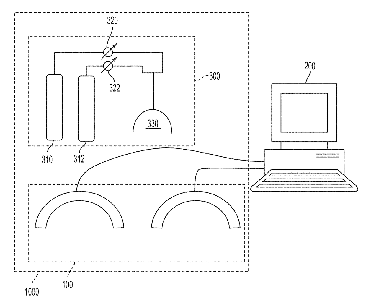
[0014]An objective measurement can be made for the detection of breast cancer with high diagnostic sensitivity and specificity, without relying on prior knowledge of whether cancer is present or which breast is affected. Novel elements of the invention leverage a combination of factors that consider how optical data is obtained from the breast, use of a simultaneous bilateral referencing protocol, and how data are treated. Specific elements disclosed here include the collection of optical array measurements from both breasts simultaneously under various protocols, including resting-state measures and evoked responses. The developed technique employs economical sensing hardware and data collection protocols that are easily implemented without patient discomfort.
[0015]To overcome the limitations imposed by the prior art, various constraints are imposed on data collection and analysis that serve to reduce the natural variance associated with optical measures of the breast. The nature of these constraints fall into three broad categories: (1) imposition of substantially symmetric external boundary conditions; (2) collection of simultaneous bilateral dynamic measures; (3) referencing measures of one breast to another. Additionally, these constraints can be applied either to data obtained under resting state conditions or to an evoked response. Results are presented demonstrating that this combination of methods serves to allow for the detection of breast cancer, and for its localization in the affected breast with high reliability, without the need for prior information.

Problems solved by technology

Favoring the use of optical methods for the detection of breast cancer, particularly in the near infrared region, are disturbances originating from tumor angiogenesis, which result in a malformed vascular bed, leading to disturbances in the hemoglobin signal.
While both static and dynamic optical measures of the breast have been implemented, there is a host of factors associated with how measures are taken, the natural variability in breast size and composition, and how data are explored, that have confounded efforts to identify useful biomarkers for the presence of breast cancer without the need for prior information.
Experience in other fields has shown that problems of this type, generally referred to as boundary value problems, can be notoriously difficult solve in any stable way.
For an appendage such as the breast, the natural variance in size, internal composition, and deformability make efforts to reliably define the boundary conditions needed to yield stable inverse solutions especially difficult.
In part because of these difficulties, efforts to apply such methods to extract biomarkers have been limited to measures of the hemoglobin signal that are time-independent.
While use of such methods does enhance the capability of distinguishing tumors from unaffected tissue, it also reduces their potential practical utility, especially if the goal is to derive biometrics that can serve as a primary screening tool.
Experience with other forms of dynamic measures (e.g., bioelectric measures of the brain or heart) indicates that there are a host of factors that can influence signal dynamics, thereby confounding interpretation of potential biomarkers.
Additionally, because these factors are uncorrelated across individuals, it can be expected that the natural variance associated with dynamics of the hemoglobin signal will be large in any group of individuals.
Such excessive variance can be expected to complicate efforts to derive useful biometrics based on dynamic measures of the breast.
An expected confounding factor here is the natural variance in breast stiffness, which would cause regions near the chest wall to experience greater loads than regions closer to the nipple.
Also confounding is the simple recognition that the signal attenuation with the optical technique is approximately a factor of ten per centimeter.
These considerations demonstrate that there are many naturally occurring factors, and elements of how data are obtained and treated, that can be expected to complicate the fidelity of derived biomarkers for the presence of cancer based on optical measures.

Method used


Image

  • Self-referencing optical measurement for breast cancer detection
  • Self-referencing optical measurement for breast cancer detection
  • Self-referencing optical measurement for breast cancer detection

Examples


embodiment

Preferred Embodiment

1. Simultaneous Bilateral Breast Measures

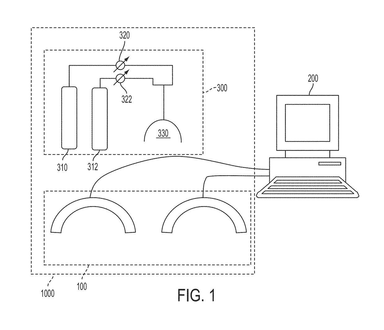
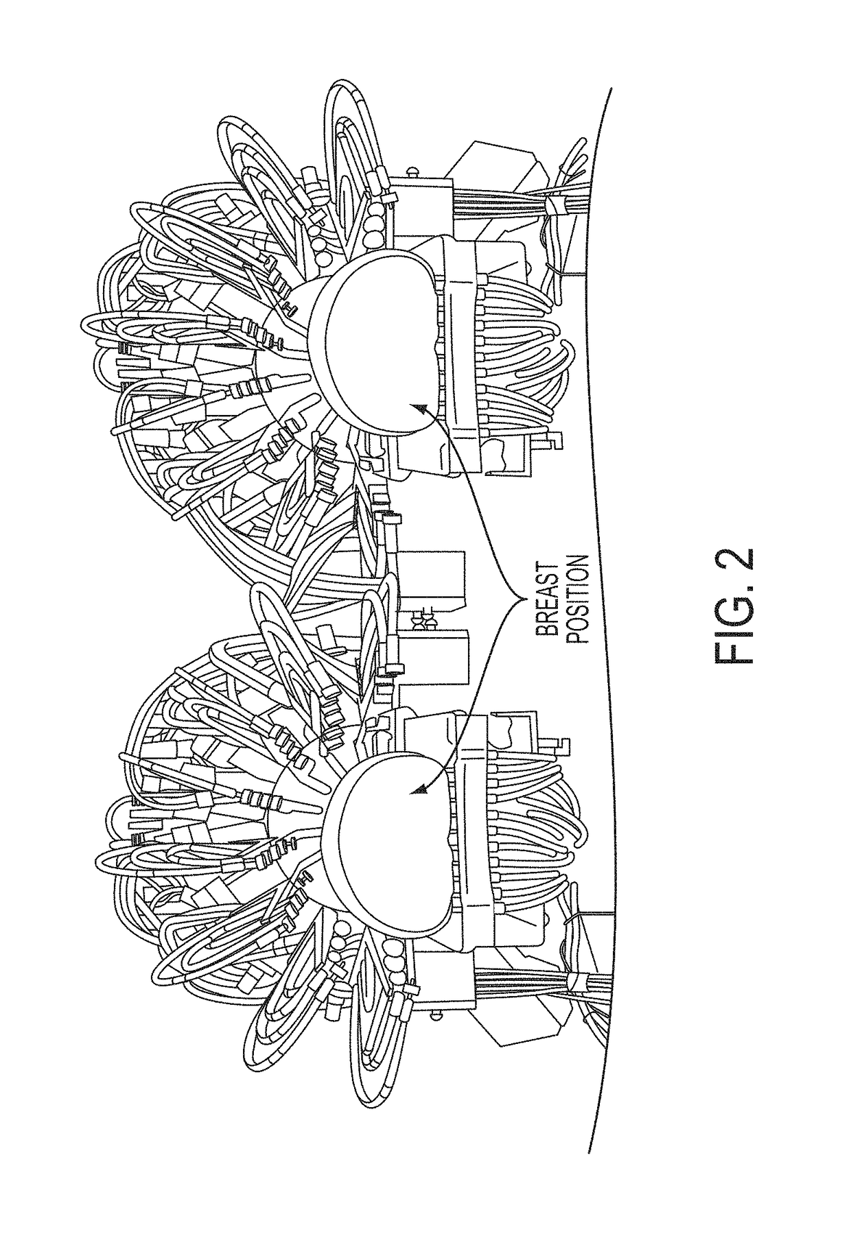
[0043]Referring to FIG. 2, a dual sensing head apparatus is shown, which has been implemented in an effort to reduce sources of variance inherent to optical measures of the breast. The dual sensing head structure (1) provides for simultaneous bilateral measures, (2) serves to impose substantially bilaterally symmetric external boundary conditions by employing feedback-controlled articulation members that can adjust optode contact force to a desired value. A description of this setup and methods used to evaluate the hemoglobin signal are given in R. Alabdi, H. L. Graber, Y. Xu, and R. L. Barbour, “Optomechanical imaging system for breast cancer detection,”J. Optical Society of America A 28, 2473-2493 (2011). Briefly, each breast is illuminated in parallel with dense array of sources (760, 830 nm), in rapid succession, with a parallel recording of the emerging light intensity.

[0044]It is appreciated by those skilled in the a...

example 1

of Breast Cancer from Resting-State Measures

[0073]In general, the resting state dynamics of the hemoglobin signal in any tissue are affected by local metabolic and systemic control mechanisms. For many forms of cancer, it is also appreciated that up-regulation of enzyme pathways accompanies the various changes in gene expression. One pathway in particular that is frequently up-regulated is that involving the inducible form of nitric oxide synthase, also known as NOS II. Among its many actions, nitric oxide (NO) is a powerful vasodilator. Previous reports in the literature have identified that subjects with breast cancer experience enhanced modulation of the spontaneous low-frequency vascular rhythms, and that this behavior is sensitive to inhibitors of NOS II. See, for example, T. M. Button, H. Li, P. Fisher, R. Rosenblatt, K. Dulaimy, S. Li, B. O'Hea, M. Salvitti, V. Geronimo, C. Geronimo, S. Jambawalikar, P. Carvelli, and R. Weiss, “Dynamic infrared imaging for the detection of ma...

example 2

of Breast Cancer from an Evoked Response

[0090]To those skilled in the art, it can be appreciated that whereas there are many forms of evoked response that could be considered that may reveal, in some instances, some type of differential response in a breast containing a tumor (e.g., use of various respiratory gas mixtures, applied force maneuvers, Valsalva maneuver) all of the concerns identified above would still apply to the expected impact that the many sources of variance would have on the stability of derived biometrics. For instance, a simple maneuver easily implemented is an applied force to achieve some level of breast compression. A brief consideration, however, indicates that the response one might observe could be highly idiosyncratic and strongly dependent on a host of external boundary and internal constituent factors (e.g., breast stiffness). In the case where the goal is to extract non-observable features (e.g., a tomographic imaging study), the obvious change in brea...


PUM


Abstract

Optical data is obtained from a pair of breasts, employing a simultaneous bilateral referencing protocol, and is subsequently analyzed employing a self-referencing data analysis method. Optical measurements can be performed on both breasts simultaneously under various protocols, including resting-state measures and evoked responses. Sensing hardware and data collection protocols are economical and can be implemented without patient discomfort. The natural variance inherently associated with optical measures of the breast is reduced by: imposition of substantially symmetric boundary conditions; collection of simultaneous bilateral dynamic measures; referencing measurement data of one breast to measurement data from another.

Description

CROSS-REFERENCE TO RELATED APPLICATIONS[0001]This application claims the benefit of U.S. Provisional Application No. 61 / 639,132, filed on Apr. 27, 2012.STATEMENT REGARDING FEDERALLY SPONSORED RESEARCH OR DEVELOPMENT[0002]This invention was made with government support under Contract No. R41CA096102 awarded by the National Institute of Health, and Contact No. DAMD017-03-C-0018 awarded by U.S. Army. The federal government of the United States has certain rights in the invention.FIELD OF THE INVENTION[0003]The present invention relates to a method for optical measurement for breast cancer detection and an apparatus for effecting the same.BACKGROUND OF THE INVENTION[0004]Diagnostic imaging methods frequently rely on evidence of anatomical disturbances that confer specific information as to the presence or absence of disease. In the case of functional techniques, such as obtained using magnetic resonance, optical or bioelectric methods, evidence of disease is based on identifying some fo...

Claims


Application Information

Patent Timeline
Patent Type & Authority Patents(United States)
IPC IPC(8): A61B6/00A61M16/10A61B10/00A61B5/00A61B5/1455
CPCA61M16/10A61B5/0073A61B5/14552A61B10/0041A61M2205/3303A61M2205/3379A61M2230/20
Inventor BARBOUR, RANDALL L.AL ABDI, RABAH M.
Owner THE RES FOUND OF STATE UNIV OF NEW YORK
Learn More