Methods and arrays for controlled manipulation of DNA and chromatin fragments for genetic and epigenetic analysis

a technology of chromatin fragments and dna, which is applied in the field of genetic and epigenetic analysis controlled manipulation of dna and chromatin fragments, can solve the problems of poor repeatability of molecule to molecule, not yet available for general use, and none of these methods is yet in general use, so as to achieve better understanding, hinder valuable information, and difficult image

Active Publication Date: 2018-01-02
CORNELL UNIVERSITY
View PDF3 Cites 0 Cited by
  • Summary
  • Abstract
  • Description
  • Claims
  • Application Information

AI Technical Summary

Benefits of technology

[0011]In another aspect, the present invention relates to a method of producing a chromatin array for use in high resolution imaging of native chromatin fragments, where this method includes: (a) providing a plurality of elongated individual native chromatin fragments removably coupled to a hydrophobic component in an orderly pattern suitable for high resolution imaging of the plurality of native chromatin fragments, where the native chromatin fragments include both DNA and histones; and (b) transferring the plurality of elongated individual native chromatin fragments to a transfer platform, thereby yielding a chromatin array including the plurality of elongated individual chromatin fragments attached to the transfer platform in said orderly pattern. The transfer platform of this method includes a support and a transfer surface layered on the support; and is effective to receive and capture the plurality of elongated individual chromatin fragments in the orderly pattern from the hydrophobic component. In one embodiment, this method further includes labeling the native chromatin fragments with at least one label for imaging or assaying purposes. The present invention also includes a chromatin array produced according to this method.
[0016]Generally, in various aspects, the present invention described herein relies on being a straightforward, low-cost and high-throughput method to controllably isolate and manipulate single chromatin fragments onto various solid supports for high-resolution imaging and analysis of multiple genetic and epigenetic marks within individual fragments.
[0018]The present invention is effective for use with various types of cells, including without limitation, mammalian and cancer cell lines in their native state. Further, the present invention is useful for high-throughput and large screening applications. In one aspect, the present invention relates to methods and devices that provide a molecular beads-on-a-string configuration that is compatible for use with high-resolution imaging and analyses using atomic force microscopy (AFM). The methods and devices of the present invention are effective for the preservation of histones during the processing steps of the present invention.
[0026]As noted herein, the present invention can be used for various applications. One particular application includes, but is not limited to, high-resolution imaging and analysis of individual chromatin fragments for genetic and epigenetic studies. This would enable a better understanding of gene regulation and disease.
[0027]The present invention also improves on aspects relating to systems, methods, and arrays described herein. As noted, conventionally, in biology, DNA is studied and characterized in its compacted state hindering valuable information. The organic composition of DNA renders it difficult to image using electron microscopy techniques that limits the use of electron beams for analysis of biological molecules. Graphene is a remarkable material that has received great interest over the past decade as it enables high contrast imaging when used as support for Scanning Electron Microscopy (SEM) or Transmission Electron Microscopy (TEM). In one embodiment, the present invention provides for the assembly of single stretched DNA molecules or chromatin fragments into regular arrays deposited on a microstructured PDMS (Polydimethylsiloxane) stamp by means of capillary assembly, and transferring this assembly from the PDMS stamp to a graphene surface (e.g., graphene TEM grids) using solvent mediation. As a result, the invention allows obtaining individual elongated molecules at predetermined locations on a graphene surface, which then enables high throughput electron beam imaging and analysis with single nucleotide resolution.
[0028]Currently, TEM imaging of DNA is generally conducted by incubating coiled DNA at the surface of a TEM grid and staining it for imagery contrast. On the contrary, the present invention provides a straightforward, low-cost and high-throughput system and method to elongate and transfer single DNA molecules (chromatin fragments, DNA-protein complexes) on graphene surfaces in one-step. This invention enables imaging of DNA with single nucleotide resolution using Electron Microscopy.

Problems solved by technology

While the benefits of single molecule analyses are known and research is underway on new such methods, they are not yet available for general use.
However, none of these methods is yet in general use in epigenetic analyses and the need for robust methods still exists.
The most popular stretching technique for FISH-like genetic or epigenetic analysis relies on cell shearing after fixation, the latter leading to poor molecule to molecule repeatability (Smith et al., Science.
Yet, to our knowledge, no method has been demonstrated that allows the controllable stretching and isolation of canonical and native chromatin molecules for high-throughput analyses while being compatible with high-resolution imaging techniques.

Method used


Image

  • Methods and arrays for controlled manipulation of DNA and chromatin fragments for genetic and epigenetic analysis
  • Methods and arrays for controlled manipulation of DNA and chromatin fragments for genetic and epigenetic analysis
  • Methods and arrays for controlled manipulation of DNA and chromatin fragments for genetic and epigenetic analysis

Examples


example 1

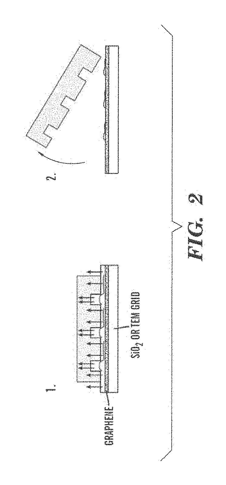
Transfer-Printing of Single DNA Molecule Arrays on Graphene for High Resolution Electron Imaging and Analysis

[0105]This example describes a new procedure for depositing ordered arrays of individual elongated DNA molecules on single-layer graphene substrates for high resolution electron beam imaging and electron energy loss spectroscopy (EELS) analysis. This demonstrates the capability to observe elemental composition of DNA with sufficient resolution to directly read genetic and epigenetic information with single base spatial resolution from individual elongated DNA molecules.

Methods

[0106]Materials.

[0107]To prepare phage lambda DNA solution, 100 μl phage lambda DNA solution (Sigma, 48 502 bp, 329 μg / ml diluted to 50 μg / ml in 10 mM Tris-HCl / 1 mM EDTA, pH 8) was heated at 65° C. for 5 min and dipped into ice water to avoid molecular concatenation. For fluorescence imaging, the solution was then fluorescently labeled with YOYO-1 intercalator (Invitrogen) by adding 1.5 μl of YOYO-1 (100...

example 2

Single DNA Molecule Patterning for High-Throughput Epigenetic Mapping

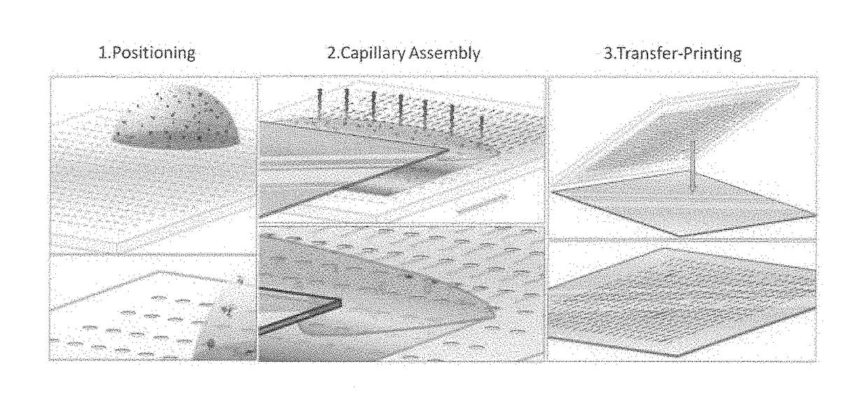
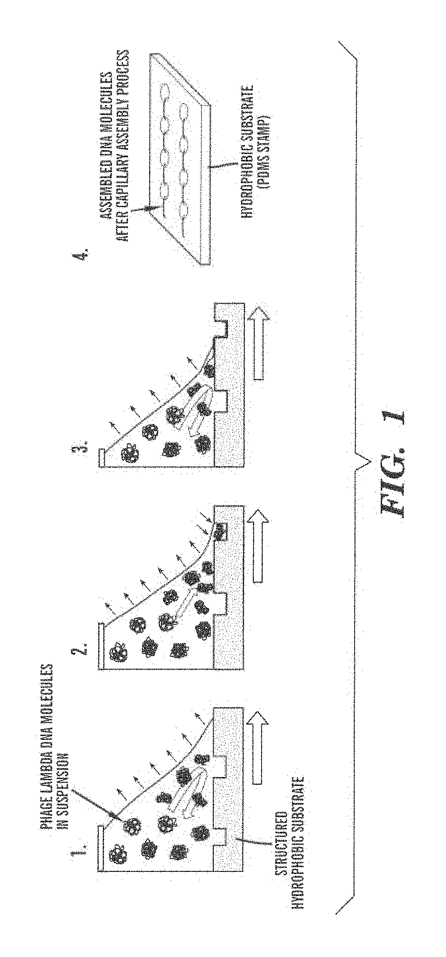
[0132]This example describes a new method for profiling the 5-methyl cytosine distribution on single DNA molecules. Our method combines soft-lithography and molecular elongation to form ordered arrays estimated to contain more than 250 000 individual DNA molecules immobilized on a solid substrate. The methylation state of the DNA is detected and mapped by binding of fluorescently labeled methyl-CpG binding domain peptides to the elongated dsDNA molecules and imaging of their distribution. The stretched molecules are fixed in their extended configuration by adsorption onto the substrate so analysis can be performed with high spatial resolution and signal averaging. We further prove this technique allows imaging of DNA molecules with different methylation states.

[0133]In this example, we present a method for transferring regular arrays of individual elongated DNA molecules onto SiO2 substrates for optical analysis. I...

example 3

Ordered Arrays of Native Chromatin Molecules for High-Resolution Imaging and Analysis

[0143]Individual chromatin molecules contain valuable genetic and epigenetic information. To date, there have not been reliable techniques available for the controlled stretching and manipulation of individual chromatin fragments for high-resolution imaging and analysis of these molecules. We report the controlled stretching of single chromatin fragments extracted from two different cancerous cell types (M091 and HeLa) characterized through fluorescence microscopy and atomic force microscopy (AFM). Our method combines soft-lithography with molecular stretching to form ordered arrays of more than 250,000 individual chromatin fragments immobilized into a beads-on-a-string structure on a solid transparent support. Using fluorescence microscopy and AFM, we verified the presence of histone proteins after the stretching and transfer process.

[0144]We present a method for the stretching of chromatin molecul...


PUM

PropertyMeasurementUnit
concentrationaaaaaaaaaa
diameteraaaaaaaaaa
diameteraaaaaaaaaa

Abstract

The present invention relates to methods and arrays for use in high resolution imaging of individual nucleic acid molecules and chromatin fragments, including native chromatin fragments. In one aspect, the present invention relates to a chromatin array that includes a transfer platform having a support and a transfer surface layered on the support. The chromatin array also includes a plurality of elongated individual native chromatin fragments coupled to the transfer surface in an orderly pattern suitable for high resolution imaging of the plurality of native chromatin fragments. The native chromatin fragments of the chromatin array include both DNA and histones.

Description

CROSS-REFERENCE TO RELATED APPLICATIONS[0001]This application is a U.S. National Phase filing under 35 U.S.C. §371 of International Application No. PCT / US2013 / 023450, filed Jan. 28, 2013, and published as WO 2013 / 112999-A1 on Aug. 1, 2013, which claims benefit of priority from U.S. Provisional Patent Application Ser. No. 61 / 591,606, filed Jan. 27, 2012, and U.S. Provisional Patent Application Ser. No. 61 / 667,491, filed Jul. 3, 2012. The entire contents of each of the prior applications are incorporated herein by reference in their entirety.GOVERNMENT RIGHTS STATEMENT[0002]This invention was made with Government support under grant number DA025722 awarded by the National Institutes of Health. The Government has certain rights in the invention.FIELD OF THE INVENTION[0003]The present invention relates to, inter alia, methods and arrays for use in high resolution imaging of individual nucleic acid molecules and chromatin fragments, including native chromatin fragments.BACKGROUND OF THE ...

Claims


Application Information

Patent Timeline
Patent Type & Authority Patents(United States)
IPC IPC(8): C12Q1/68G01N1/30
CPCC12Q1/6806C12Q1/6837G01N1/30C12Q2522/101
Inventor CERF, ALINECRAIGHEAD, HAROLD G.TIAN, HARVEY C.
Owner CORNELL UNIVERSITY
Why Eureka
Try Eureka