Production of amniotic extractive liquid and use thereof

A technology of extract and amniotic membrane, which is applied in the field of preparation of amniotic membrane extract, can solve the problems of negative serological examination during the incubation period, and achieve the effects of avoiding visual impact, reducing risk, and being easy to store

Inactive Publication Date: 2009-07-15
ZHONGSHAN OPHTHALMIC CENT SUN YAT SEN UNIV
View PDF3 Cites 0 Cited by
  • Summary
  • Abstract
  • Description
  • Claims
  • Application Information

AI Technical Summary

Problems solved by technology

However, there are false-negative results in serological tests, or pregnant women have been infected with the virus, but they are in the window period of virus infection when the serological test is done (if they are infected with the virus but are still in

Method used


Image

  • Production of amniotic extractive liquid and use thereof
  • Production of amniotic extractive liquid and use thereof

Examples


Embodiment 1

[0028] Example 1: Preparation of Amnion Extract

[0029] The prenatal serological diagnosis was negative for HIV, hepatitis B virus, hepatitis C virus, and syphilis, and the placenta with normal fetal development was selected for elective cesarean section and brought back quickly on ice under sterile conditions.

[0030] Rinse twice with pre-cooled medical saline on the ultra-clean bench according to the sterile standard of cell culture to remove residual blood, then soak the placenta in medical saline containing 50u / ml penicillin and 50mg / ml streptomycin, The amniotic membrane was separated from the chorion starting from the amniotic membrane tear, and the residual chorionic tissue on the basal surface of the amniotic membrane was removed with a small piece of dry sterile medical gauze.

[0031] After grinding with liquid nitrogen, add pre-cooled medical saline at a weight-to-volume ratio of 1:10. The amniotic membrane is fully homogenized by a mechanical homogenizer on the i...

Embodiment 2

[0034] Embodiment 2: Amniotic membrane extract is used in the experiment of animal model

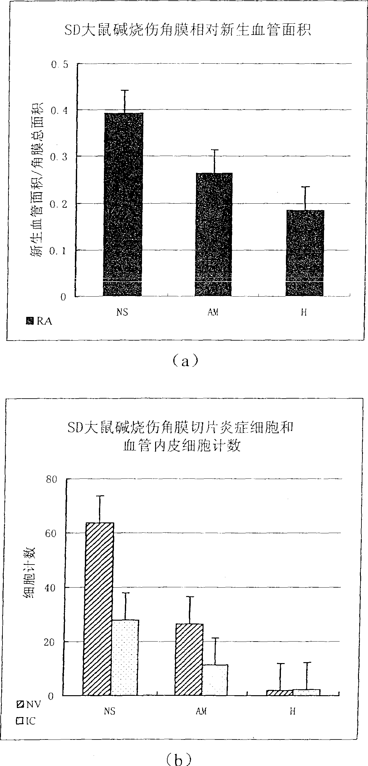
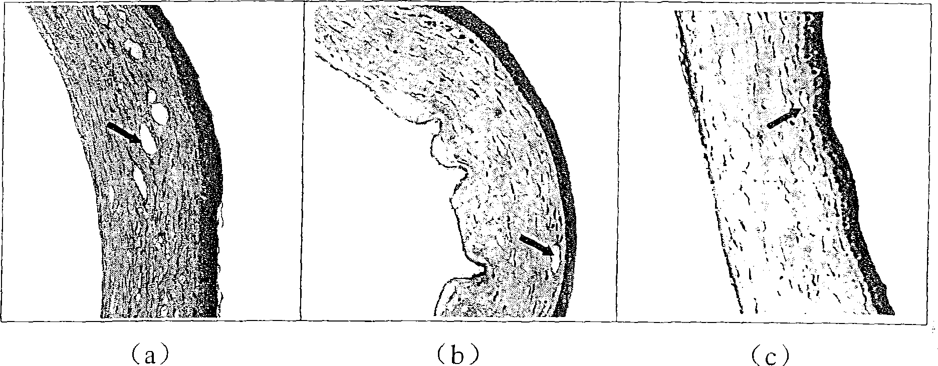
[0035] The eye drops prepared from the amniotic membrane extract were used to treat corneal alkali burns in animal models, and it was found that the eye drops had the effects of inhibiting new blood vessels and inhibiting inflammation. The specific experimental process is as follows:

[0036]Take 30 SD rats, burn the corneas of both eyes of the rats with 1M NaOH for 15 seconds, and the burning scar is located in the center of the cornea with a diameter of 3mm. Eight hours after instillation of tobramycin eye ointment, they were randomly divided into amniotic membrane extract group, normal saline control group and hormone control group, with 10 eyes in each group, 20 eyes in total. (NS) and compound dexamethasone eye drops (H) (prepared by Zhongshan Ophthalmic Center of Sun Yat-sen University, containing 0.1% dexamethasone sodium and 0.5% neomycin sulfate) eye drops, four times a day (8a...


PUM


Abstract

The invention relates to a preparation method of amniotic membrane extract. The preparation method comprises the following steps: taking the cesarean section placenta, separating the amniotic membrane after washing, grinding with liquid nitrogen, adding medical physiological saline according to the weight-to-volume ratio of 1:5-10, and homogenizing to obtain a homogenate; the homogenate is placed at 0-4°C 1-7 days, heat at 100-120°C for 30-60 minutes, filter, and take the filtrate; adjust the pH of the filtrate to 3, let it stand, filter, and take the filtrate; adjust the pH of the filtrate to 7.2, 0.20-0.22 under sterile conditions um membrane filter, take the filtrate, which is the placental tissue extract, and store at 0-4°C. The preparation method of the invention has a simple process, can obtain a relatively high-concentration extract, is easy to store, is safe to use, and is suitable for clinical application. The present invention also relates to the use of the amnion extract for preparing medicines for inhibiting ocular surface neovascularization and inflammatory diseases, especially for the preparation of medicines for treating corneal alkali burns.

Description

technical field [0001] The invention relates to a preparation method of amnion extract and a new application of amnion extract. Background technique [0002] The amniotic membrane is a layer of membranous tissue attached to the placenta, separated from the placenta only by a layer of chorion. The placenta is rich in blood vessels, but the amnion is a transparent tissue. The clinical record of amniotic membrane was first seen in 1910. At first, the amniotic membrane and chorion were transplanted at the same time. In 1935, the amniotic membrane was separated separately for transplantation. In terms of the preparation and application of amniotic membrane, the First People's Hospital of Hefei once applied for the Chinese invention patent "Preparation Method of Human Amniotic Membrane" in 1997. Application, promotes wound healing and reduces tendon adhesions. In ophthalmology, the preparation of amniotic membrane and its clinical application were patented by Tseng in the Unite...

Claims


Application Information

Patent Timeline
IPC IPC(8): A61K35/50A61P27/02
Inventor 刘祖国蒋爱华
Owner ZHONGSHAN OPHTHALMIC CENT SUN YAT SEN UNIV
Why Eureka
Try Eureka