Eureka AIR delivers breakthrough ideas for toughest innovation challenges, trusted by R&D personnel around the world.

Blood type method system and device

A technology for blood and red blood cells, applied in measurement devices, biochemical cleaning devices, biochemical equipment and methods, etc., can solve the problems of not allowing cross-matching, large test tubes, false negative results, etc., and achieve fast and accurate results. The effect of obtaining results, low error tolerance

Inactive Publication Date: 2008-02-06
INVERNESS SWITZERLAND GMBH
View PDF5 Cites 4 Cited by
  • Summary
  • Abstract
  • Description
  • Claims
  • Application Information

AI Technical Summary

Problems solved by technology

Disadvantages of this method include: (a) several minutes of mixing required; (b) subjective visual judgment of results; (c) false positive responses due to drying; and (d) slides cannot be stored
Disadvantages of the tube agglutination method include: (a) the need for laboratory equipment, personnel, and environment; (b) subjective visual judgment of results; (c) incorrect centrifugation speed or time may lead to false positive results or false negative results , because cell clumps may be interpreted as immunoagglutination; (d) no direct recording of results; (e) bulky tubes; and (f) risk of breakage and spillage
Disadvantages are: (a) need for experimental equipment, personnel, and environment; (b) no direct result recording (c) bulky test hardware (although the manufacturer refers to the product as a "card", it is actually located on a support series of test tubes and must not be as flat as a "card"); and (d) a complex manufacturing process (inserting the gel into the narrow tube) which results in high costs
Although the Akers device simplifies blood grouping to the point that it can be used by non-blood bank professionals outside the laboratory (e.g., at the bedside), it has certain disadvantages: (a) it requires a relatively large amount of blood; (b) it requires Operator timing of steps; (c) it only allows forward blood grouping and does not allow cross-matching (ability to visualize recipient antibodies against donor red blood cells); (d) yields potentially misleading results: used in Results The absence of red in the window indicates a positive result and the presence of a red color indicates a negative reaction; and (e) the result must be observed exactly 1 minute after adding the saline wash
While these products do simplify blood testing procedures to some extent, they are not completely error-free and can be complicated for some personnel (Rachel and Plapp, 1990; Migeot et al., 2002)

Method used


Image

  • Blood type method system and device
  • Blood type method system and device

Examples


example

[0150] The examples that follow are exemplary implementations of methods and systems according to the invention. These examples are not intended to be limiting in any way.

[0151] Example 1 - Lotion

[0152] The washes used to generate the instances are:

[0153] A. Dulbecco's Phosphate Buffered Saline (PBS) obtained from Biological Industries, Beit Ha'emek, Israel.

[0154] B. A solution prepared from PBS diluted 1:1 in water with 4% w / v polyethylene glycol (PEG) 15000-20000 MW (Fluka) and 0.3% w / v dextran sulfate sodium salt (Amersham Biosciences).

[0155] C. Dulbecco's Phosphate Buffered Saline (PBS) with 0.001-0.01% w / v polyoxyethylene-10-tridecyl ether (Sigma).

example 2

[0156] Example 2 - Blood Grouping

[0157] Ahlstrom #142 filter paper of size 0.5 x 0.5 cm was placed on the absorbent pad. Two μL of whole blood was pipetted into the center of the filter followed by 2 μL of anti-blood typing reagent (Gamma Biologicals Inc., Houston, Texas, USA). The filter was then rinsed with a few drops (from the dropper bottle) of wash solution and dried. Alternatively, use Ahlstrom #142 filter paper with a 4mm circle radius, 10 μL of blood, and 10 μL of antiblood group reagent, followed by a few drops of the wash solution of Example 1.

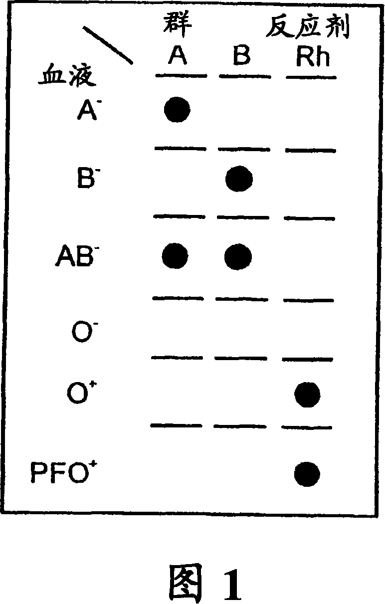
[0158] This test was repeated with various blood samples of different blood types (received from and pre-tested by the Blood Service Center of Israel RedMagen David, Tel Hashomer, Israel) and tested with anti-A, anti-B, anti-AB and anti-D (=Rh ) blood group reagent for testing. A - Blood only produces red spots with anti-A and anti-AB reagents. B - Blood was spotted with anti-B and anti-AB reagents. AB - Blood ...

example 3

[0159] Example 3 - Grouping of Blood Using Dried Reagents

[0160] Ahlstrom #142 filter discs of size 0.5 x 0.5 cm were placed on a non-absorbent surface (hollow bottom, disposable Petri dish). 50 [mu]L of anti-blood typing reagent (Gamma Biologicals, Inc., Houston, TX, USA) was pipetted onto each piece of filter paper. Petri dishes were incubated overnight at 37°C with filter paper containing anti-blood typing reagents. At this point the filter is dry.

[0161] To test the ability of the dried antibody-dipped discs to correctly identify blood groupings of whole blood samples, they were placed on absorbent pads and 1 μL of test blood was placed approximately in the center of each filter pad of the filter pad series. The series comprising pads are each impregnated with one of anti-A, anti-B or anti-D (=Rh) reagents.

[0162] Rinse remaining blood from the filter pad by placing 5-10 drops of wash solution A or B. The filters are then air dried for record keeping. The resu...


PUM


Abstract

A method for the detection and / or visualization of particle agglutination in a particle suspension is provided, comprising placing a volume of the particle suspension and a volume of a solution or suspension containing an agglutinating agent at substantially the same selected location on a surface of a filter constructed so as to permit passage of individual unagglutinated particles in a direction perpendicular to the surface. A wash solution at optionally placed at substantially the same location as that of said agglutinating agent, and the surface is observed for the presence of particles. There is also provided a method for detection of agglutination reactions, such as used in blood grouping, reverse grouping and cross-matching. A device and a kit based on the invention is also claimed, which facilitates blood grouping, reverse grouping and matching in non-laboratory environment without the need for laboratory instruments.

Description

technical field [0001] The present invention relates generally to the detection of receptor-ligand interactions, and more particularly to the detection of blood group antigens and antibodies thereto for forward and reverse blood typing and matching purposes as employed in transfusion medicine. Background technique [0002] Due to its simplicity, rapidity and relative sensitivity, particle agglutination is a widely used immunological method for the detection and visualization of antigen-antibody interactions (Riochet 1993). Cells in general, and red blood cells in particular, are particles amenable to various methods of agglutination. Likewise, agglutination of red blood cells (hemagglutination) is used in blood typing or matching as an important part of the pre-transfusion testing process to detect antigens on their surface and antibodies to these antigens (Rouger 1993; Brecher 2002). The purpose of the pre-transfusion test of blood is to prevent adverse reactions caused by...

Claims


Application Information

Patent Timeline
Patent Type & Authority Applications(China)
IPC IPC(8): G01N33/00G01N33/567C12M1/34C12Q1/56
CPCG01N33/5302G01N33/543G01N33/80G01N33/54313
Inventor G·罗特F·塞缪尔尔斯F·菲什A·本-兹维
Owner INVERNESS SWITZERLAND GMBH
Why Eureka
Eureka Blog
Learn More