Apatite developer and its positron emitting tomography imaging
A technology of apatite and developer, applied in the field of biomedical materials, can solve problems such as difficulty in obtaining three-dimensional dynamic distribution and metabolic images, and difficult technical operations
- Summary
- Abstract
- Description
- Claims
- Application Information
AI Technical Summary
Problems solved by technology
Method used
Image
Examples
Embodiment 1
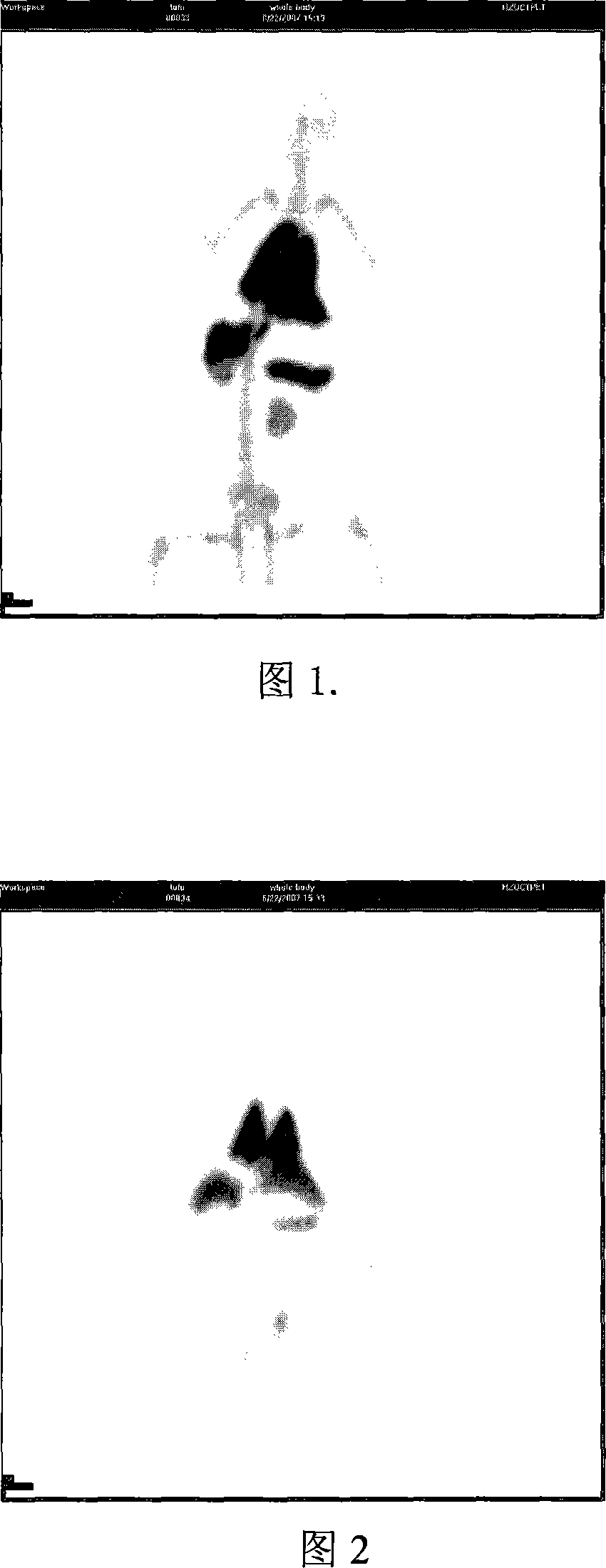
[0034] Prepare the same volume of 0.01 mol / L calcium hydroxide aqueous solution and 0.006 mol / L phosphoric acid aqueous solution, adjust the calcium hydroxide aqueous solution to pH 10 with ammonia water, and then slowly add the phosphoric acid aqueous solution to the calcium hydroxide aqueous solution through a constant flow pump During the dropwise addition, the oil bath was maintained at 90°C. After the dropwise addition, the positron nuclides generated by the cyclotron 18 F (or 76 Br, 75 Br, 124 1) Pour into the mixed solution, continue to maintain the stirring state, and keep the temperature at 60°C. After 2.5 hours, the suspension containing apatite developer was taken out, washed three times with deionized water, and the measured specific activity was 0.014mCi / mg.
[0035] One healthy New Zealand white rabbit was taken, and after being fully anesthetized, apatite contrast agent injection with a radiation dose of 1.02mCi was injected through the ear vein. Immediately...
Embodiment 2
[0038] Prepare the same volume of 1 mol / liter calcium acetate monohydrate aqueous solution and 0.6 mol / liter ammonium dihydrogen phosphate aqueous solution, adjust the calcium acetate monohydrate to pH 7.0 with ammonia water, and prepare the calcium and fluorine molar ratio of 10:0.01 in diphosphate Add ammonium fluoride into the ammonium hydrogen solution, and then slowly drop the ammonium dihydrogen phosphate solution dissolved in ammonium fluoride into the calcium acetate monohydrate solution through a constant flow pump, and keep the oil bath at 80°C. Accelerator-produced positron nuclides 18 Pour F into the mixture, keep stirring and keep the temperature at 60°C. After 3 hours, the suspension containing apatite developer was taken out, washed 3 times with deionized water, and the measured specific activity was 0.134mCi / mg.
[0039] Two healthy New Zealand white rabbits were taken, and after being fully anesthetized, 1.01 mCi of apatite buffered saline dispersion was inje...
Embodiment 3
[0041] Prepare the same volume of 0.5 mol / liter calcium chloride aqueous solution and 0.3 mol / liter diammonium hydrogen phosphate aqueous solution, adjust the calcium chloride aqueous solution to a pH value of 8.5 with ammonia water, and prepare the calcium chloride aqueous solution according to the calcium-zinc molar ratio of 10:0.5 in calcium chloride Zinc chloride is added to the aqueous solution, and ammonium fluoride is added to the diammonium hydrogen phosphate aqueous solution according to the molar ratio of calcium to fluorine of 10:1, and then the diammonium hydrogen phosphate aqueous solution is slowly added dropwise to the calcium chloride aqueous solution through a constant flow pump to maintain oil bath at 60°C, the positron nuclides produced by the cyclotron 18 Pour F into the mixture, keep stirring and keep the temperature at 60°C. After 3 hours, the suspension containing apatite developer was taken out, washed 5 times with deionized water, and the measured spec...
PUM
 Abstract
Description
Claims
Application Information
 - R&D
- Intellectual Property
- Life Sciences
- Materials
- Tech Scout
- Unparalleled Data Quality
- Higher Quality Content
- 60% Fewer Hallucinations
Browse by: Latest US Patents, China's latest patents, Technical Efficacy Thesaurus, Application Domain, Technology Topic, Popular Technical Reports.
© 2025 PatSnap. All rights reserved.Legal|Privacy policy|Modern Slavery Act Transparency Statement|Sitemap|About US| Contact US: help@patsnap.com

