Method for placenta mesenchyma stem cell separation and in vitro amplify cultivation

A technology for mesenchymal stem cells and in vitro expansion, applied in biochemical equipment and methods, animal cells, tissue culture, etc., can solve the problem of unclear specific marker molecules of placental mesenchymal stem cells, low subculture and expansion ability, It is difficult to form mesenchymal stem cell lines and other problems, and achieve the effect of being conducive to clinical safe transplantation application, improving expansion ability, and obvious expansion advantages

Inactive Publication Date: 2008-09-24
ZHEJIANG UNIV
View PDF0 Cites 69 Cited by
  • Summary
  • Abstract
  • Description
  • Claims
  • Application Information

AI Technical Summary

Problems solved by technology

The subculture and expansion ability of this multicellular mixture is low, and it is difficult to form a true mesenchymal stem cell line
Although the specific marker molecules

Method used


Image

  • Method for placenta mesenchyma stem cell separation and in vitro amplify cultivation
  • Method for placenta mesenchyma stem cell separation and in vitro amplify cultivation
  • Method for placenta mesenchyma stem cell separation and in vitro amplify cultivation

Examples


Embodiment 1

[0021] A specific method that the method of the present invention takes is: get placental maternal side decidua, cut into pieces fetal decidua side placental tissue, collect cells after 0.1% type IV collagenase digestion, use cell culture medium (containing DMEM, 10% fetal bovine serum, 2mML-glutamine) for adherent culture.

[0022] 1. After the cells are layered in the culture flask, discard the culture medium and suspended cells. Adherent spindle cells were digested with Tryspin-EDTA and harvested. Harvested cells were washed once with PBS and suspended in PBS. Then, according to the instructions provided by MACS, CD34 was sorted from the suspension cells with the adsorption CD14 monoclonal antibody-magnetic bead separation system (MACS, Miltenyi Biotech Inc., Shanghai). - cell. collected CD34 - After the cells were cultured on the wall again, the cells were harvested, washed once with PBS, and then CD34 was sorted using the adsorption CD105 monoclonal antibody-magnetic ...

Embodiment 2

[0025] Separation and purification efficiency

[0026] The proportions of different labeled cell surface antigens in cells obtained by different separation and purification techniques were measured by flow cytometry, as shown in Table 1.

[0027] Table 1 The proportion (%) of different labeled cell surface antigens in cells obtained by different separation and purification techniques

[0028]

[0029] CD34 in primary placental maternal decidua cells + , CD45 + , CD105 + and CD29 + The average cells were 7.22%, 11.65%, 47.14% and 34.62%, indicating that the primary adherent spindle cells were dominated by mesenchymal cells. After 2 passages of adherent culture, CD34 + Cells and CD45+ cells decreased, while CD90+ and CD105 cells increased, but also contained a small amount of CD34 + cells and CD45+ cells. After CD14 monoclonal antibody and CD105 monoclonal antibody-magnetic bead system sorting, the obtained cells are basically CD34 - CD45 - CD105 + CD29 + cell. Ac...

Embodiment 3

[0031] Cell morphology obtained by different sorting methods

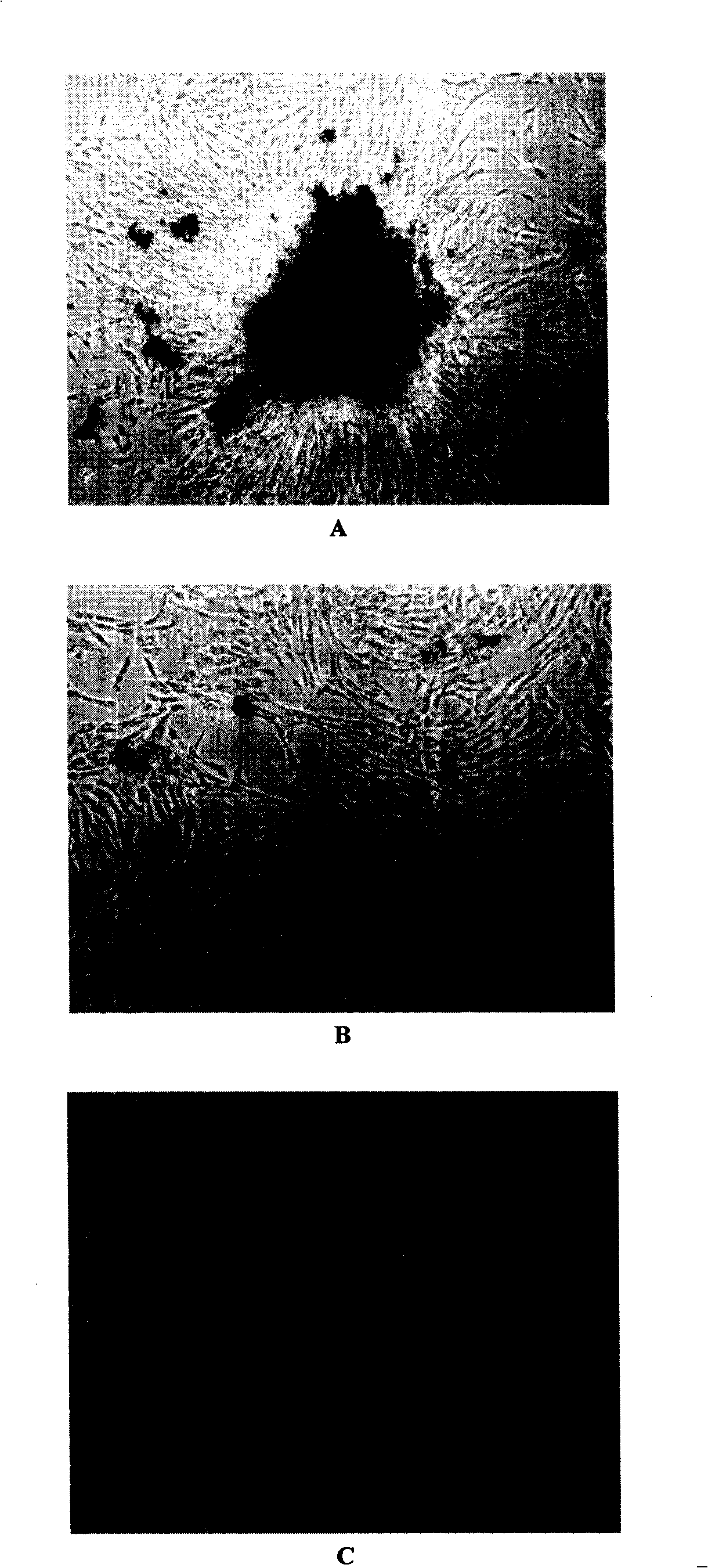
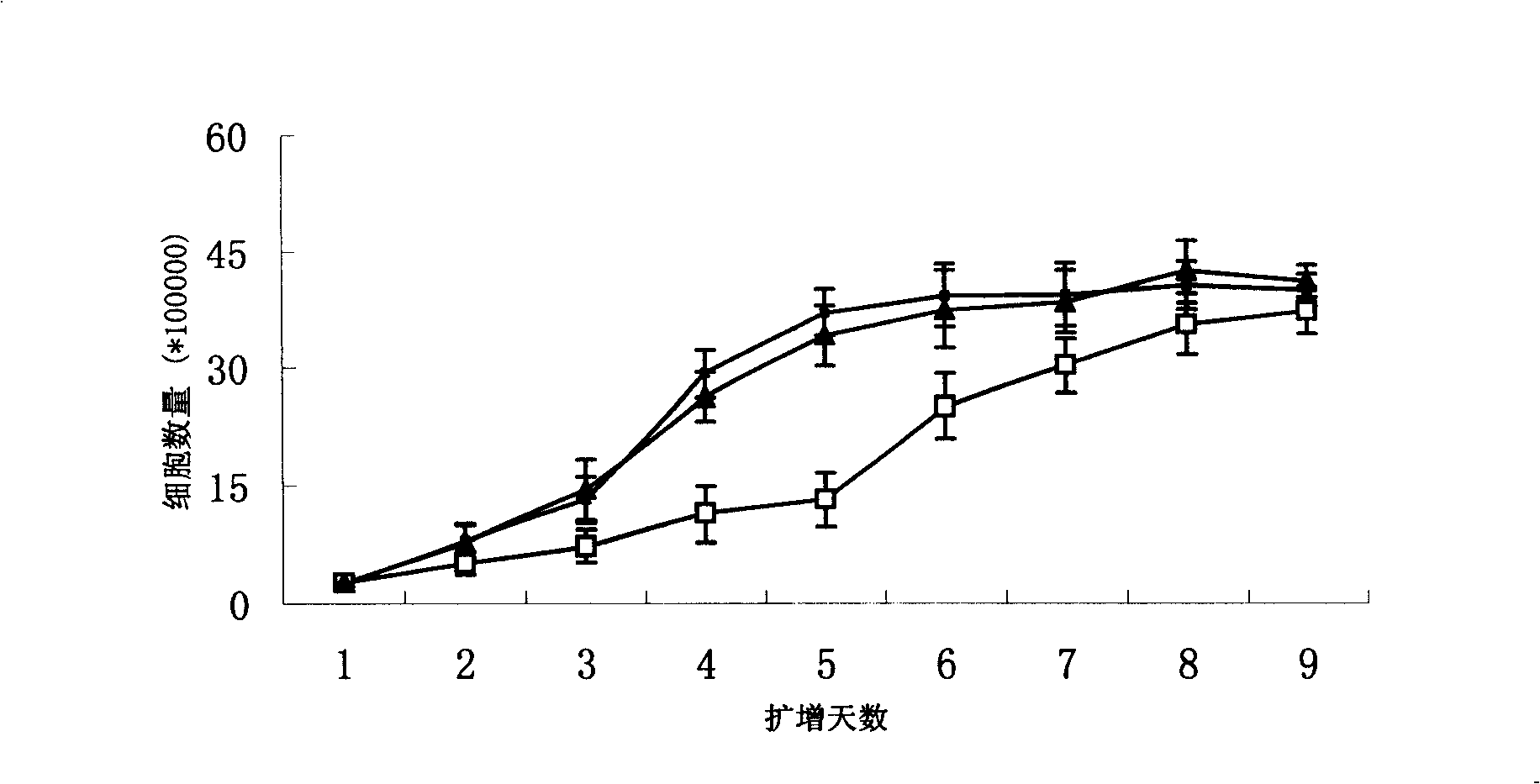
[0032] see figure 1 24 hours after inoculation, long spindle-shaped cells could be seen in placental tissue cells digested with type IV collagenase and arranged radially or concentrically. A large number of colonies appeared 4 days after inoculation and gradually layered on the bottom of the culture flask. It takes 14-16 days for cell expansion to form a monolayer in Teflen-25 flasks. figure 1 A shows that the cells are spindle-shaped under an inverted microscope, 10×10 magnifications. After the cells were subcultured twice, the cells showed remarkable fibrous cells, and a cell monolayer was formed after 8-9 days after cell inoculation ( figure 1 B). Placental mesenchymal stem cells (CD34 - CD105 +) under a new serum-free culture system (ginsenoside polysaccharide / DMEM-LG / 10% FBS) to carry out the first-generation expansion culture, and form a cell monolayer in a Teflen-25 culture flask after 5-6 days. figur...


PUM


Abstract

The invention provides a method for separation of placenta-derived mesenchymal stem cells, and for amplification and culture in vitro. After decidual tissue cells on the side of a placenta matrix are adopted and collected, primary culture cells are amplified by adhering to wall; the placenta-derived mesenchymal stem cells are purified by adopting positive and negative immunological sorting and combination method to get CD34<->CD105<+> cells which are secondarily amplified and cultured in a serum-free culture system in vitro. By adopting the method provided by the invention, just few primary culture cells (1X10<5> cells) are needed every time to produce a good sorting result; and the purification rate of the placenta-derived mesenchymal stem cells is further improved. Not only the culture system used has significant advantage of amplification over placenta-derived mesenchymal stem cells, but also the amplified cells have multiplex differentiation potential. The method can be applied for purification of placenta-derived mesenchymal stem cells and amplification and culture in vitro.

Description

Technical field [0001] The invention belongs to biological technology, and relates to a method for isolating placental mesenchymal stem cells and expanding and culturing in vitro. This method can effectively purify placental mesenchymal stem cells and improve their ability to expand in vitro. Background technique [0002] A type of mesenchymal stem cells with multi-differentiation potential exists in human term placenta. Related studies have shown that this kind of stem cells can differentiate into osteoblasts, chondrocytes, fat cells and nerve cells. This kind of stem cells is called placental mesenchymal stem cells (MSCs), which will be of great value in cell transplantation and tissue engineering in medicine. [0003] Since no specific marker molecule for placental mesenchymal stem cells has been found yet, the isolation and purification of the stem cells is still an indirect method: the traditional method is to flush out the placental residual blood through the umbilic...

Claims


Application Information

Patent Timeline
IPC IPC(8): C12N5/08G01N33/53C12N5/0775
Inventor 王金福袁文佶石东燕
Owner ZHEJIANG UNIV
Why Eureka
Try Eureka