Method for preparing cellulose contrast medium for stomach and intestine ultrasonic imaging

An ultrasound contrast agent, cellulose technology, applied in echo/ultrasonic imaging agents, etc., can solve the problems of short residence time, unfavorable inspection, uneven particle size, etc., and achieve the effect of eliminating artifacts

Inactive Publication Date: 2008-10-08
李丽君
View PDF0 Cites 3 Cited by
  • Summary
  • Abstract
  • Description
  • Claims
  • Application Information

AI Technical Summary

Problems solved by technology

At the same time, this type of silent contrast agent often cannot form an obvious contrast effect with gastrointestinal lesions and the hierarchical structure of the gastric wall. In addition, this type of contrast agent is very easy to produce a rear echo enhancement effect, which can image the posterior wall of the gastrointestinal tract and surrounding tissues and organs. make to make (2) There are echo-type acoustic contrast agents: main...

Method used


Image

  • Method for preparing cellulose contrast medium for stomach and intestine ultrasonic imaging
  • Method for preparing cellulose contrast medium for stomach and intestine ultrasonic imaging
  • Method for preparing cellulose contrast medium for stomach and intestine ultrasonic imaging

Examples


preparation example Construction

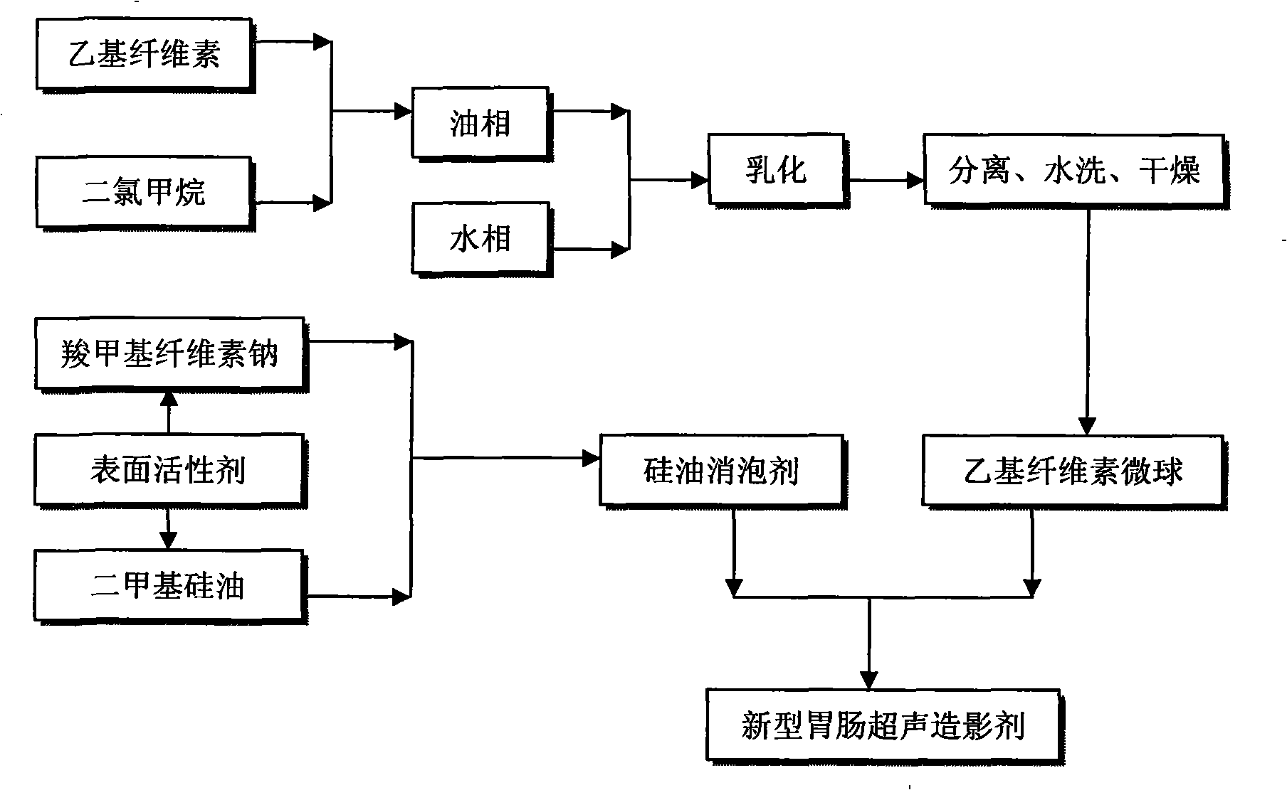
[0045] Preparation of ethylcellulose microspheres:

[0046] (1) Weighing ethyl cellulose and dissolving it in dichloromethane to prepare an organic phase with a concentration of 10-30% by mass;

[0047] (2) Weigh polyvinyl alcohol and dissolve it in distilled water to prepare an external aqueous phase solution with a concentration of 1 to 5% by mass;

[0048] (3) Add the organic phase dropwise to the external aqueous phase solution at a speed of 300-1500r / min, stir and disperse for 5-30min;

[0049] (4) After the solvent is volatilized for 2 to 8 hours at a stirring speed of 500 to 600 r / min, centrifuge and wash with water for 3 to 5 times, and dry to obtain ethyl cellulose microspheres with a particle size of 20 μm;

[0050] Preparation of silicone oil defoamer:

[0051] The silicone oil defoamer refers to medical, edible grade silicone oil and other medical, edible grade surfactants. The preparation method is as follows:

[0052] (1) Weigh carboxymethylcellulose sodium, add...

Embodiment 1

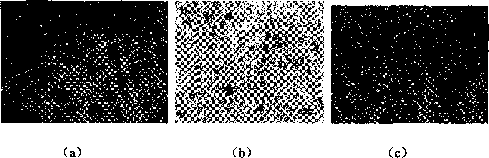
[0065] Weigh 1.0 g of ethyl cellulose and dissolve it in 10 ml of dichloromethane solution as the organic phase. At the same time, 3.0 g of polyvinyl alcohol (PVA-20) was weighed and added to 100 ml of distilled water as the external aqueous phase solution, and 2 g of Tween-60 was added thereto, and heated to dissolve. The organic phase solution was added dropwise to the external aqueous phase at a rotational speed of 800r / min, stirred for 10min, and then moved to a magnetic stirrer at a stirring speed of 500r / min, and the solvent was evaporated for 5h. The microspheres were separated by centrifugation, washed three times with distilled water, and then dried to obtain ethylcellulose microspheres after repeated three times. Observed by an optical microscope, the surface of the microspheres is smooth without adhesion, and the particle size is relatively concentrated, with an average particle size of 23.2 μm. When preparing the silicone oil defoamer, first weigh 0.5g sodium carb...

Embodiment 2

[0067] Weigh 1.0 g of ethyl cellulose and dissolve it in 15 ml of dichloromethane solution as the organic phase. At the same time, 3.0 g of polyvinyl alcohol (PVA-20) was weighed and added to 100 ml of distilled water as the external aqueous phase solution, and 0.5 g of Tween-60 and 0.5 g of Tween-80 were added thereto, and heated to dissolve. The organic phase solution was added dropwise to the external aqueous phase at a rotational speed of 1200r / min, stirred for 10min, and then moved to a magnetic stirrer at a stirring speed of 550r / min, and the solvent was evaporated for 5h. The microspheres were separated by centrifugation, washed with distilled water, repeated three times, and dried to obtain ethylcellulose microspheres. Observed by an optical microscope, the surface of the microspheres is smooth without adhesion, and the particle size is relatively concentrated, with an average particle size of 18.6 μm. When preparing the silicone oil defoamer, first weigh 0.5g sodium ...


PUM

PropertyMeasurementUnit
Particle sizeaaaaaaaaaa
The average particle sizeaaaaaaaaaa

Abstract

The invention discloses a method for preparing a cellulose gastrointestinal ultrasound angiography agent. Ethyl cellulose microspheres are firstly prepared; then a silicon oil defoamer is prepared; finally, the ethyl cellulose microspheres with a grain diameter of 20 microns and the silicon oil defoamer are mixed according to mass proportion of 1 to 100; thus the cellulose gastrointestinal ultrasound angiography agent is obtained. Due to the ability of eliminating gastrointestinal gases, the silicon oil defoamer can effectively eliminate artifacts caused by the interference of gases; evenness of a development image is determined by the grain diameter and the distribution of the ethyl cellulose microspheres; when the grain diameter is 20 microns and the distribution of the granularity is even, the ultrasound development effect is the best. The angiography agent of the invention not only ensures clear display of the structure of gastrointestinal wall, but helps to clearly distinguish surrounding organs and is expected to be used in clinical examination of gastrointestinal power.

Description

technical field [0001] The invention belongs to the technical field of medical ultrasonic diagnosis, and relates to a preparation method of a cellulose gastrointestinal ultrasonic contrast agent. Background technique [0002] Gastrointestinal contrast-enhanced ultrasonography, also known as gastric filling method, uses a contrast agent to fill the stomach and intestinal cavity to eliminate the interference of gas in the stomach and intestinal cavity on ultrasound, so as to improve the gastrointestinal ultrasound environment and create a new environment for ultrasound examination. A good acoustic window not only enables the normal gastrointestinal wall structure and lesions to be clearly displayed, but also distinguishes the surrounding organs more clearly, and the contrast agent itself does not affect the penetration of sound waves. Therefore, the performance of the contrast agent directly affects the image quality and diagnostic accuracy. [0003] Traditionally, ultrasonog...

Claims


Application Information

Patent Timeline
IPC IPC(8): A61K49/22
Inventor 李丽君
Owner 李丽君
Why Eureka
Try Eureka