Pharmaceutical for promoting functional regeneration of damaged tissue

A tissue regeneration and cell technology, applied in tissue culture, antipyretics, animal cells, etc., can solve the problems of poor prognosis of life, insufficient effect of immune-gene therapy, etc., and achieve the effect of promoting functional tissue regeneration

Inactive Publication Date: 2009-02-25
GENOMIX CO LTD +1
View PDF1 Cites 22 Cited by
  • Summary
  • Abstract
  • Description
  • Claims
  • Application Information

AI Technical Summary

Problems solved by technology

In addition, the life prognosis of malignant glioma is also poor, and immuno-gene therapy, which has been widely studied in recent years, including chemotherapy and radiation therapy, has not shown sufficient effect

Method used


Image

  • Pharmaceutical for promoting functional regeneration of damaged tissue
  • Pharmaceutical for promoting functional regeneration of damaged tissue
  • Pharmaceutical for promoting functional regeneration of damaged tissue

Examples


Embodiment 1

[0259] Objective: To evaluate the role of bone marrow-derived cells in functional regeneration of skin tissue transplanted in living organisms

[0260] Method: For the above purpose, research was carried out by the following method.

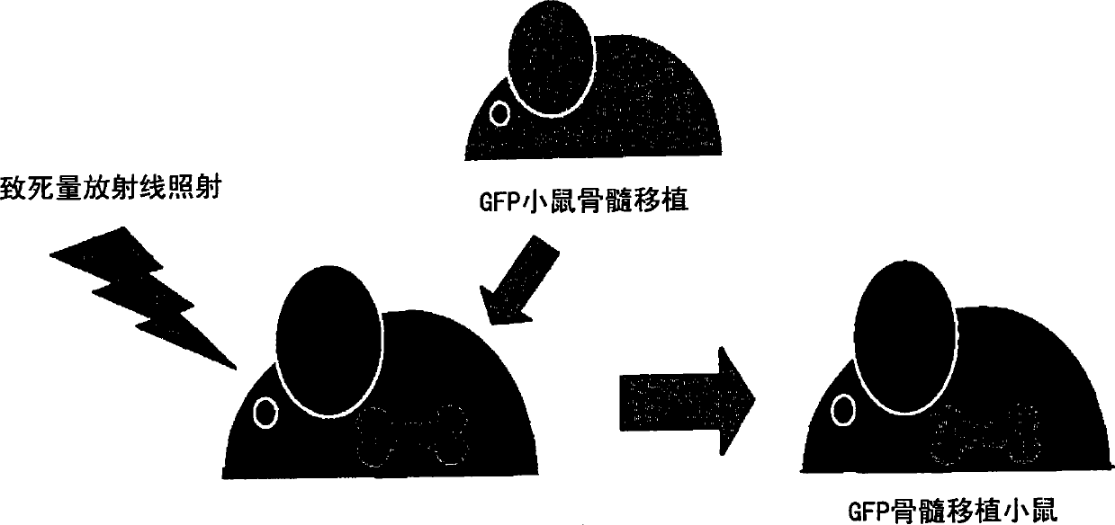
[0261] 1) The degree of contribution of bone marrow-derived cells to the functional regeneration of the grafted skin sheet was investigated using a living skin graft system to GFP bone marrow transplanted mice. Specifically, C57BL / 6 male mice (6-8 weeks old) were irradiated with lethal radiation (10 Gy), and immediately thereafter transplanted GFP (green fluorescent protein) transgenic mouse-derived bone marrow cells (5×10 6 Each / 0.1ml physiological phosphate buffer solution pH7.4)( figure 1 ).

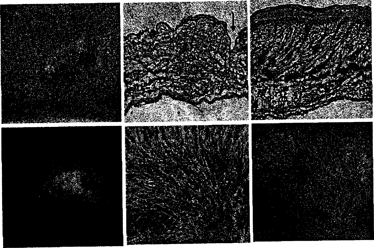
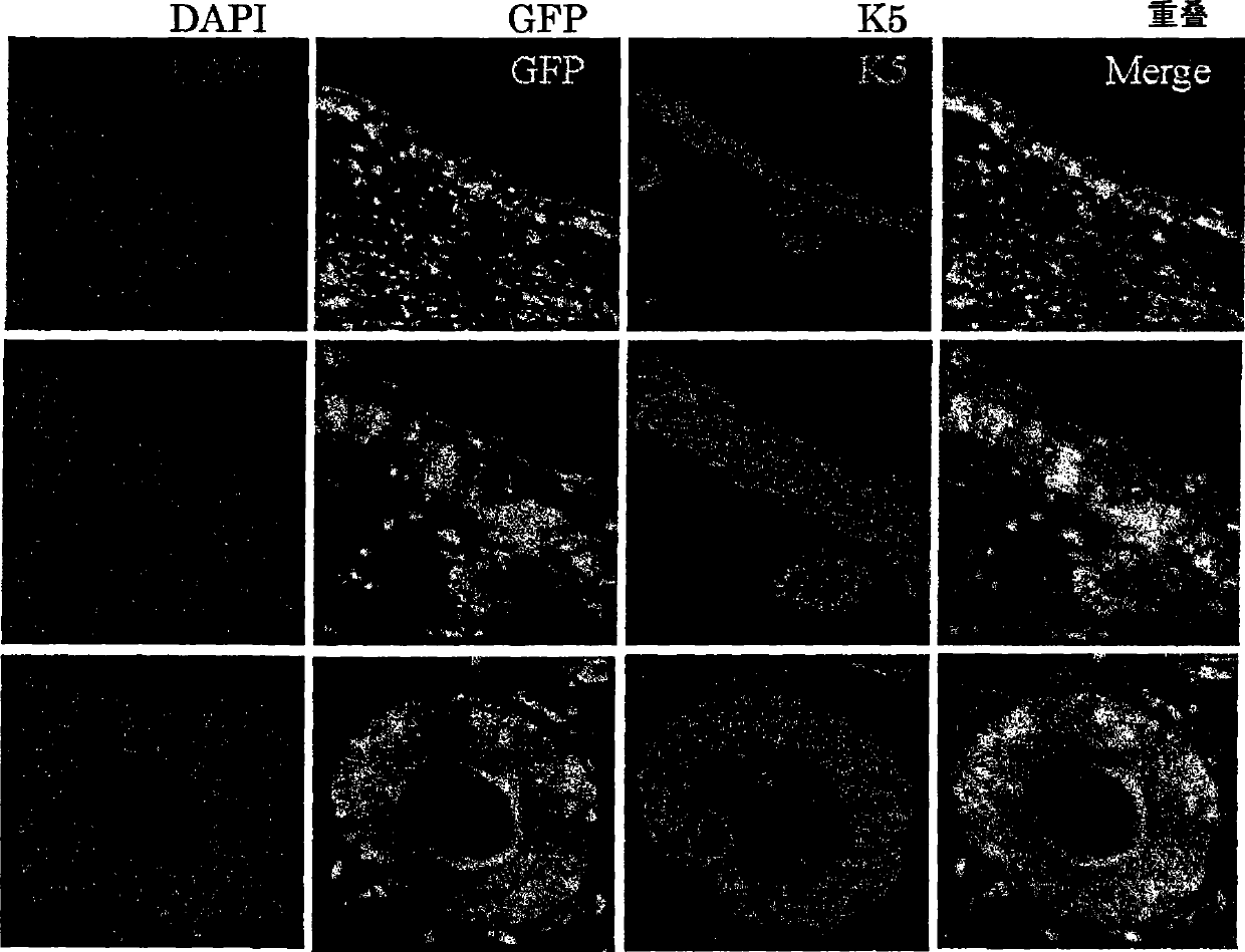
[0262] 2) Waiting for the engraftment of transplanted bone marrow cells (6 weeks), newborn mouse skin (female) was transplanted on the back skin of the obtained GFP bone marrow transplanted mouse.

[0263] 3) Waiting for the implantation of the transp...

Embodiment 2

[0269] Objective: Identification of bone marrow mesenchymal stem cell-inducing factors present in skin tissue extracts

[0270] Method: In order to identify the bone marrow mesenchymal stem cell mobilization factor expected to be released from the resected skin in a state of hypoperfusion, the following method was used for the study.

[0271] 1) To obtain mouse bone marrow-derived mesenchymal stem cells, bone marrow cells of C57BL / 6 mice were collected from the femur or lower leg bone, and D-MEM (manufactured by Nacalai Co., Ltd.) containing 10% fetal bovine serum was used as a cell culture medium. Inoculated in a cell culture dish and cultured at 37° C. and a carbon dioxide concentration of 5%. When the area occupied by the cells grew to 70 to 100% of the bottom area of ​​the culture dish, the cells were detached from the culture dish using 0.25% trypsin 1 mM EDTA (manufactured by Nacalai), and then subcultured under the same conditions. The subculture operation was repeated...

Embodiment 3

[0283] Objective: To identify the HMGB1 family in skin extracts and study the induction activity of bone marrow mesenchymal stem cells

[0284] Method: The presence or absence of the HMGB protein family contained in the skin extract of newborn mice was confirmed by Western blot. As a sample, 10 μl of the skin extract obtained in [Example 2] was used for electrophoresis by SDS-PAGE, and the protein separated in the gel was transferred to a PVDF membrane using a blotting apparatus (ATTO). After incubating at room temperature for 1 hour with PBS (S-T-PBS) containing 3% skimmed milk and 0.1% Tween 20, the rabbit anti-mouse HMGB1 antibody, rabbit anti-mouse HMGB2 antibody, rabbit anti-mouse HMGB2 antibody, and rabbit Anti-mouse HMGB3 antibodies were reacted at 4°C for 16 hours. After the reaction, wash the same PVDF membrane with S-T-PBS for 5 minutes and 5 times, and then use peroxidase-labeled goat anti-rabbit IgG antibody (GE Healthcare) diluted to 2000 times with S-T-PBS, and ...


PUM


Abstract

The present inventors revealed for the first time in the world that 1) a large amount of bone marrow-derived cells are mobilized in transplanted skin; 2) the mobilized bone marrow-derived cells are differentiated into any of dermal fibroblasts, adipocytes, muscle cells, vascular endothelial cells and epidermal keratinocytes in a transplanted skin graft, and in the mobilized bone marrow-derived cells, bone marrow-derived mesenchymal stem cells are contained; 3) what mobilize the bone marrow-derived mesenchymal stem cells to the graft from the peripheral blood are HMGB1, HMGB2 and HMGB3 released from necrotic tissue of the transplanted skin; 4) purified HMGB1, HMGB2 and HMGB3 promote migration of mesenchymal stem cells isolated from the bone marrow and cultured; 5) active substances including HMGB1 that allow bone marrow mesenchymal stem cells to migrate can be simply purified from an extract of a large number of organs including skin, brain and heart; 6) active substances that allow bone marrow mesenchymal stem cells to migrate can be simply extracted from cultured cells; and 7) a heparin column-purified fraction of skin extract mobilizes a large amount of bone marrow-derived cells during brain damage.

Description

technical field [0001] The present invention relates to a drug for promoting function regeneration of damaged tissue. Background technique [0002] In recent years, the contribution of various stem cells to the repair process of damaged tissues has been clarified, and the development of new regenerative medicine that induces functional tissue regeneration by mobilizing a large number of stem cells to the damaged site has been actively developed. In order to realize this new type of regenerative medicine, it is necessary that: 1) there are abundant stem cells that can be mobilized to the damaged site in the living body, and 2) the factors that mobilize the stem cells to the damaged site can be isolated and identified. [0003] Stem cells that can be mobilized to the injury site include tissue stem cells present in the injury site or nearby tissues and bone marrow-derived stem cells present in peripheral blood. In recent years, it has been reported that bone marrow-derived ce...

Claims


Application Information

Patent Timeline
Patent Type & Authority Applications(China)
IPC IPC(8): A61K38/00A61K31/711A61K35/12A61K35/76A61K48/00A61P1/04A61P9/10A61P11/00A61P17/00A61P17/02A61P17/14A61P19/08A61P29/00A61P37/06A61P43/00C07K14/47C12N5/06C12N15/00C12Q1/00G01N33/15G01N33/50A61K35/28A61K35/36C07K14/46C12N5/071C12N5/077C12N5/0775C12N15/09
CPCG01N2500/10A61K31/711A61K38/00G01N33/5073C07K14/52G01N33/6863A61P1/04A61P11/00A61P17/00A61P17/02A61P17/14A61P19/08A61P29/00A61P37/06A61P43/00A61P9/10A61K38/17A61K48/00
Inventor 玉井克人山崎尊彦金田安史
Owner GENOMIX CO LTD
Why Eureka
Try Eureka