Tissue engineered bone-cartilage complex tissue graft and preparation method thereof

A technology of composite tissue and tissue engineering, applied in medical science, prosthesis, etc., can solve the problems of poor quality of cartilage part and poor combination of osteochondral interface, etc., and achieve the effect of increasing adhesion ability

Inactive Publication Date: 2009-05-27
JINAN UNIVERSITY
View PDF0 Cites 17 Cited by
  • Summary
  • Abstract
  • Description
  • Claims
  • Application Information

AI Technical Summary

Problems solved by technology

The osteochondral complex constructed by this method has poor cartilage quality, mostly fibrocartilage and less hyaline cartilage. At the same time, the degree of integration of the complex depends on the maturity of each component, regardless of in vivo or in vitro experiments. , the constructed osteochondral interface is poorly combined

Method used

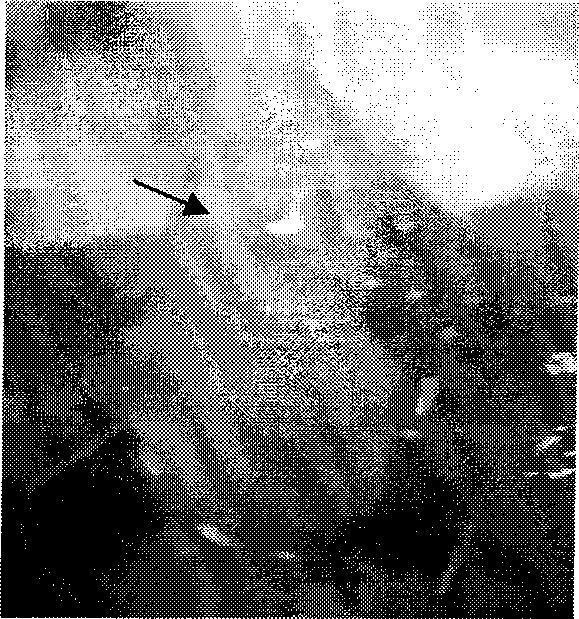
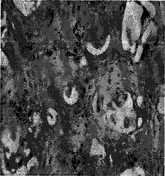
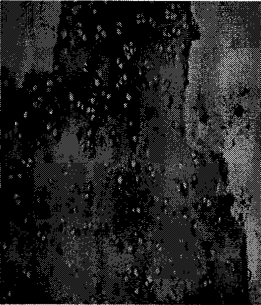

Image

  • Tissue engineered bone-cartilage complex tissue graft and preparation method thereof
  • Tissue engineered bone-cartilage complex tissue graft and preparation method thereof
  • Tissue engineered bone-cartilage complex tissue graft and preparation method thereof

Examples


Embodiment 1

[0031] 1. In vitro culture of BMSCs

[0032] One-month-old SD rats were killed by dislocation of the neck, and the bilateral femurs were removed, and the soft tissues were removed, soaked in 75% alcohol for 5 minutes, washed twice with PBS, cut off one end of the femur, and the volume fraction was 10% with a syringe with a No. 5 needle. Wash the bone marrow cavity of the femur with DMEM (containing 100 U / ml penicillin, 100 μg / ml streptomycin, and 20 U / ml heparin) of fetal bovine serum, and slowly inject 4 ml of the mixed bone marrow fluid into the pre-prepared centrifuge tube containing 6 ml of lymphocyte separation medium , centrifuge at 800G for 20min, take out the mononuclear cells in the middle layer and inoculate them on 25cm 2 Culture flask, 37°C, 5% CO 2 Culture in an incubator. Change the medium for the first time after 5 days, and change the medium every 3 days thereafter. Passage after overgrown, and take the third generation of cells for experiments.

[0033] 2. ...

Embodiment 2

[0038] As another preferred embodiment, the difference between this embodiment and embodiment 1 is:

[0039] In the synthesis steps of PLGA preparation, in PBS solution 1, the concentration of bFGF was 1 μg / ml; in PBS solution 2, the concentration of TGF-β1 was 1 μg / ml; in PBS solution 3, the concentration of BMP-2 was 1 μg / ml.

[0040] The pore diameter of the osteogenic part of the scaffold material is 50 μm, and the porosity is 85%; the pore diameter of the chondrogenic part of the scaffold material is 200 μm, and the porosity is 95%.

Embodiment 3

[0042] As another preferred embodiment, the difference between this embodiment and embodiment 1 is:

[0043] In the synthesis steps of PLGA preparation, in PBS solution 1, the concentration of bFGF was 3 μg / ml; in PBS solution 2, the concentration of TGF-β1 was 3 μg / ml; in PBS solution 3, the concentration of BMP-2 was 3 μg / ml.

[0044] The pore diameter of the osteogenic part of the scaffold material is 80 μm, and the porosity is 88%; the pore diameter of the chondrogenic part of the scaffold material is 180 μm, and the porosity is 92%.


PUM

PropertyMeasurementUnit
concentrationaaaaaaaaaa
pore sizeaaaaaaaaaa
pore sizeaaaaaaaaaa

Abstract

The invention provides a tissue engineered bone-cartilage composite tissue transplant and a preparation method thereof, and belongs to the tissue engineering field. The method comprises the following steps: seed cells are implanted on a scaffold material and cultured in vitro to form the tissue engineered bone-cartilage composite tissue transplant; the seed cells are osteogenic stem cells; the scaffold material comprises an osteogenic part and a chondroblast part, and the osteogenic part is added with an osteoblast induction factor and modified by a type-I collagen in a preparation process; and the chondroblast part is added with a chondroblast induction factor and modified by a type-II collagen. Bone marrow mesenchymal stem cells are successfully induced into osteocytes and chondrocytes, and bone tissues and cartilage tissues are formed after the obtained transplant is transplanted in vivo. Satisfactory effect is obtained from the tissue engineered bone-cartilage composite tissue which is constructed by the transplant.

Description

technical field [0001] The invention belongs to the technical field of tissue engineering, and in particular relates to a tissue-engineered bone-cartilage composite tissue graft and a preparation method thereof. Background technique [0002] Tissue engineering technology is to plant a small amount of autologous cells on the scaffold material after in vitro culture and expansion, and then construct a cell-scaffold complex similar to the shape, structure and biological characteristics of the lesion under the action of biological factors, so as to repair and Replacing damaged tissue, it has advantages that traditional treatment methods cannot match, and is considered to be the next commanding height that needs to be challenged in regenerative medicine. [0003] The repair and reconstruction of bone and joint injuries is a long-term clinical problem that is difficult to solve, especially the repair of combined bone and cartilage injuries in joints is even more difficult. Mature...

Claims


Application Information

Patent Timeline
Patent Type & Authority Applications(China)
IPC IPC(8): A61L27/38A61L27/18A61L27/54
Inventor 查振刚林宏生吴昊
Owner JINAN UNIVERSITY
Why Eureka
Try Eureka