Perultimate chromomere marker of cotton chromosome fluorescent in situ hybridization and use thereof

A fluorescence in situ hybridization and chromosome technology, applied in the field of molecular cytogenetics, can solve the problems of inability to separate and obtain telomere sequences, difficulties in cloning telomere sequences, and difficulty in inserting telomere sequences

Inactive Publication Date: 2009-07-08
INST OF COTTON RES CHINESE ACAD OF AGRI SCI
View PDF0 Cites 5 Cited by
  • Summary
  • Abstract
  • Description
  • Claims
  • Application Information

AI Technical Summary

Problems solved by technology

[0005] Direct cloning of telomere sequences is very difficult. Guo Ge et al. (1996) did microdissection of chromosomes, and Chen Shouyi et al. (2003) failed to clone telomere sequences through telomere-related sequences.
This is because the telomere at the end of the chromosome is composed of linear DNA molecules. Usually, one of the two strands of DNA paired with each other is slightly longer than the other, forming a t-loop structure. The existence of the t-loop makes the te

Method used


Image

  • Perultimate chromomere marker of cotton chromosome fluorescent in situ hybridization and use thereof
  • Perultimate chromomere marker of cotton chromosome fluorescent in situ hybridization and use thereof
  • Perultimate chromomere marker of cotton chromosome fluorescent in situ hybridization and use thereof

Examples


Embodiment 1

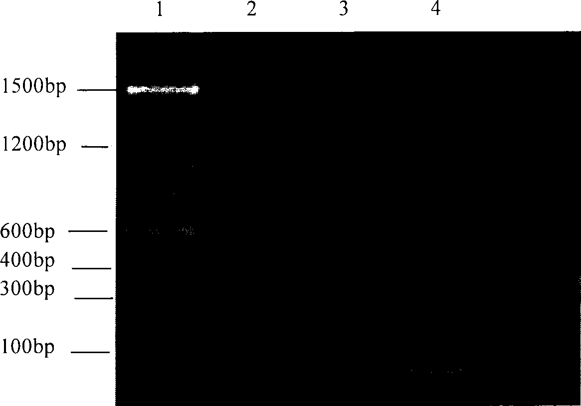
[0022] Example 1 Cloning of Telomere Sequence

[0023] By searching the nucleic acid sequences published on NCBI, it was found that a BAC clone (F23H6) located on chromosome 3 of Arabidopsis thaliana contained a segment of Arabidopsis thaliana telomere sequence. A pair of primers were designed according to the non-tandem repeat sequence at both ends of the telomeric sequence: TS1 (5'-TGAGGGACTCACATACATTG-3'); TS2 (5'-ACCACTGAACATCCATTGAG-3'). The Arabidopsis genome was used as a template for PCR amplification. The conditions of the PCR reaction are: the reaction system is first denatured at 94°C for 5 minutes, followed by 30 cycles, each cycle consists of the following reaction conditions: denaturation at 94°C for 45s, annealing at 55°C for 30s, extension at 72°C for 45s, The final step was an extension at 72°C for 5 min. The product of the PCR reaction was purified by gel recovery and ligated with T-easy carrier. The obtained clone containing the target fragment was named ...

Embodiment 2

[0024] Example 2 Telomere markers of diploid (2n=2x=26) cotton chromosome fluorescence in situ hybridization:

[0025] Material: African cotton (Cotton Research Technology and Trade Company, Chinese Academy of Agricultural Sciences)

[0026] 1. Preparation of root tip metaphase chromosomes:

[0027]Take dozens of African cotton seeds, use a scalpel to cut the seed shell at the seed growth point, and then sow the seeds in boiled and sterilized moist fine sand, and germinate at 25-30°C. When the root grows to 1-2cm (about 1 and a half days), take the root tip (0.5mm) and pretreat it with cycloheximide (25ppm) at 20°C (to break the spindle silk, so that the prophase chromosomes can be concentrated in advance and form dispersed metaphase chromosome), pretreatment for 2 hours in spring and autumn, 1.5 hours in summer, and 2.5 hours in winter. After pretreatment, wash with distilled water for 10 minutes, and fix with fixative solution (95% ethanol: glacial acetic acid 3:1) for 2-2...

Embodiment 3

[0066] Example 3 Telomere markers of tetraploid (4n=4x=52) cotton chromosome fluorescence in situ hybridization:

[0067] Material: Tetraploid cotton Xinhai 7# (Cotton Research Technology and Trade Company, Chinese Academy of Agricultural Sciences)

[0068] 1. Preparation of root tip metaphase chromosomes

[0069] Soak Xinhai 7# seeds in warm water at about 60°C overnight, sow in boiled and sterilized moist fine sand, and germinate at 25-30°C. When the root grows to 3-5cm (about 2 and a half days), take the root tip (0.5mm), and pretreat it with cycloheximide (25ppm) at 20°C (to break the spindle silk, so that the prophase chromosomes can be concentrated in advance to form dispersed metaphase chromosome), pretreatment for 2 hours in spring and autumn, 1.5 hours in summer, and 2.5 hours in winter. After pretreatment, wash with distilled water for 10 minutes, and fix with fixative solution (95% ethanol: glacial acetic acid 3:1) for 2-24 hours. Then, wash with water for 10min,...


PUM


Abstract

The present invention provides a telomere marker suitable for cotton fluorescent in situ hybridization, which is a sequence marked by biotin shown in the SEQ ID No.1. The telomere marker is taken as probe and cotton root tip somatic cell metaphase chromosome to perform fluorescence in situ hybridization (FISH), and stronger hybridization signals occur at the end of the chromosome. Further experiments confirm that the in situ hybridization signal is located at the extreme end of chromosome, that is, the hybridization signal is the telomere signal. Therefore, the length of the cotton somatic cell metaphase chromosome can be estimated by the telomere marker located at the end of chromosome, so as to lay the foundation for further karyotype analysis.

Description

technical field [0001] The invention belongs to the field of molecular cytogenetics, and in particular relates to a terminal telomere marker which can be used for fluorescence in situ hybridization of cotton chromosomes and an application thereof. Background technique [0002] DNA fluorescence in situ hybridization (FISH) technology is an important non-radioactive in situ hybridization technology developed in the late 1970s and early 1980s. Its basic principle is to mark the DNA probe with a specially modified nucleotide molecule (such as biotin-dUTP or digoxigenin-dUTP), and hybridize with the corresponding sequence on the chromosome according to the principle of DNA-DNA or DNA-RNA base pairing. When performing in situ hybridization, the DNA molecule is first treated with high temperature or alkali to denature it; when the temperature drops or the pH value returns to neutral, the denatured DNA will re-form hydrogen bonds according to the principle of complementary base pair...

Claims


Application Information

Patent Timeline
IPC IPC(8): C12Q1/68C12N15/11C12P19/34
Inventor 凌键王坤波王春英宋国立刘方黎绍惠张香娣王玉红
Owner INST OF COTTON RES CHINESE ACAD OF AGRI SCI
Why Eureka
Try Eureka