Protein suspending chip for quantitative detection of staphylococcal enterotoxin B and method for producing the same

A Staphylococcus intestinalis, suspension chip technology, applied in measuring devices, material analysis by observing the influence of chemical indicators, instruments, etc., can solve problems such as interference inspection results, and achieve high sensitivity and wide dynamic detection range Effect

Inactive Publication Date: 2009-08-05
CHINESE ACAD OF INSPECTION & QUARANTINE
View PDF0 Cites 5 Cited by
  • Summary
  • Abstract
  • Description
  • Claims
  • Application Information

AI Technical Summary

Problems solved by technology

0.01ng staphylococcal enterotoxin can be detected, but if the food leachate is impure, it can often interfere with the test results

Method used


Image

  • Protein suspending chip for quantitative detection of staphylococcal enterotoxin B and method for producing the same
  • Protein suspending chip for quantitative detection of staphylococcal enterotoxin B and method for producing the same
  • Protein suspending chip for quantitative detection of staphylococcal enterotoxin B and method for producing the same

Examples


preparation example Construction

[0044] 2. Preparation of samples to be tested

[0045] 1. Sample preparation

[0046] The target detection sample is staphylococcal enterotoxin B (SEB). Interfering samples or samples used as method-specific tests are other bacteria or other proteins other than the target detection object, including BONT, recombinant HIV P24 antigen, BSA, casein, tryptone, avian influenza virus HA protein, NH protein, etc. The above-mentioned samples to be analyzed were homogeneously dissolved in the sample diluent PB (0.01M, pH7.2) solution, and stored at 4°C. The stock solution concentration of SEB was 1mg / mL. Toxin and recombinant protein samples were diluted just before use. The concentration range of SEB was 10 pg / mL-5 μg / mL. In comparative experiments, the same samples were used for ELISA and suspension chip detection.

[0047] The bacteria to be analyzed were diluted into 10-fold different gradients with PB, and the staphylococcal enterotoxin was diluted into 4-fold different gradi...

Embodiment 1

[0052] Example 1, capture antibody-coated encoded microspheres

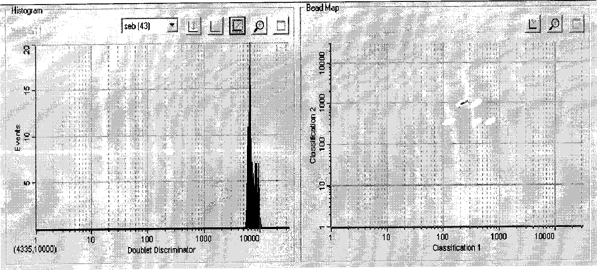
[0053] Rabbit anti-SEB was purified by n-octanoic acid-saturated ammonium sulfate purification. Optionally, one of the encoded microspheres (such as 043) is labeled with an SEB-capturing antibody (SEB antibody).

[0054] A. Activation of encoded microspheres

[0055] Take 100 μL of encoded microspheres into a 1.5 mL centrifuge tube, centrifuge at 14,000 g, carefully aspirate and discard the supernatant. Add 100 μL of microsphere washing buffer to suspend, shake and sonicate, centrifuge at 14000g, carefully aspirate and discard the supernatant. Add 100 μL of microsphere activation buffer, then add 10 μL of freshly prepared EDC (50 mg / mL), then add 10 μL of freshly prepared Sulfo-NHS (50 mg / mL), and shake at room temperature for 20 min. Add 150 μL of PBS (pH7.4), shake, centrifuge at 14000 g, carefully aspirate and discard the supernatant. Add 100 μL of PBS (pH 7.4) to suspend the encoded microspheres.

[0056...

Embodiment 2

[0060] Example 2, Biotin labeling of detection antibody

[0061] Antibodies to be labeled include rabbit anti-SEB antibody, goat anti-SEB antibody, and SEB monoclonal antibody. There are at least two options for detection antibodies labeled with biotin for each analyte. Prepare 10mM biotin solution and 2mg / mL antibody solution to be labeled respectively, add the calculated volume of biotin to the antibody solution to be labeled, shake at room temperature for 30min (or 2 hours on ice), and pass through the column for desalting Aliquot and store at -20°C for later use.


PUM


Abstract

The invention aims to provide a protein suspension chip capable of quantificationally detecting bacterial toxin and the preparation method thereof, and particularly relates to a protein suspension chip which is suitable for quantificationally detecting and analyzing staphylococcal enterotoxin. The method has high sensitivity and high particularity, good detection capacity and wide dynamic range and builds a novel detection modularity platform.

Description

technical field [0001] The invention relates to a protein suspension chip for quantitative detection of staphylococcal enterotoxin B and a preparation method thereof. Background technique [0002] Staphylococcal enterotoxin B (SEB) is the main pathogenic factor causing food poisoning. The onset of food poisoning is more acute, usually within 1-6 hours of eating toxin-containing food, with an average of 2-3 hours. [0003] SEB conventional laboratory diagnostic methods include: 1) Immunodiffusion: Oudin one-way diffusion technique can be used for accurate quantitative determination of staphylococcal enterotoxin, which is easy to repeat and is not affected by temperature, antiserum dilution and action time. The sensitivity of detecting various types of staphylococcal enterotoxins is about 10-20 μg / mL. 2) Radioimmunoassay (RIA): It is 100 times more sensitive than the immunodiffusion method for detecting enterotoxins A and B in food. 3) Reverse Passive Hemagglutination Assay...

Claims


Application Information

Patent Timeline
Patent Type & Authority Applications(China)
IPC IPC(8): G01N33/546G01N21/78G01N33/569
Inventor 王静姜永强孙肖红杨宇胡孔新谢士嘉
Owner CHINESE ACAD OF INSPECTION & QUARANTINE
Why Eureka
Try Eureka