In vivo micro-invasive investigation device including a metal guide

A guiding device and equipment technology, applied in the direction of catheter, medical science, diagnosis, etc., can solve the problems of correct direct access to the target site, correct guidance of damage, complex use, etc., to achieve the effect of easy navigation, improved flexibility or elasticity

Active Publication Date: 2014-05-07
BIOSTEMS LTD
View PDF7 Cites 1 Cited by
  • Summary
  • Abstract
  • Description
  • Claims
  • Application Information

AI Technical Summary

Problems solved by technology

The latter would then fall into too complex a use to properly reach its target site without simultaneously changing the path of the blood circulation
At the same time, with regard to the acquisition of the rigidity required to perforate the organ or tissue, connecting the various elements impairs the overall flexibility of the device and thus its correct guidance

Method used


Image

  • In vivo micro-invasive investigation device including a metal guide
  • In vivo micro-invasive investigation device including a metal guide
  • In vivo micro-invasive investigation device including a metal guide

Examples


Embodiment 1

[0113] Example 1: Manufacture and activation of metal guides based on nickel-titanium memory alloys

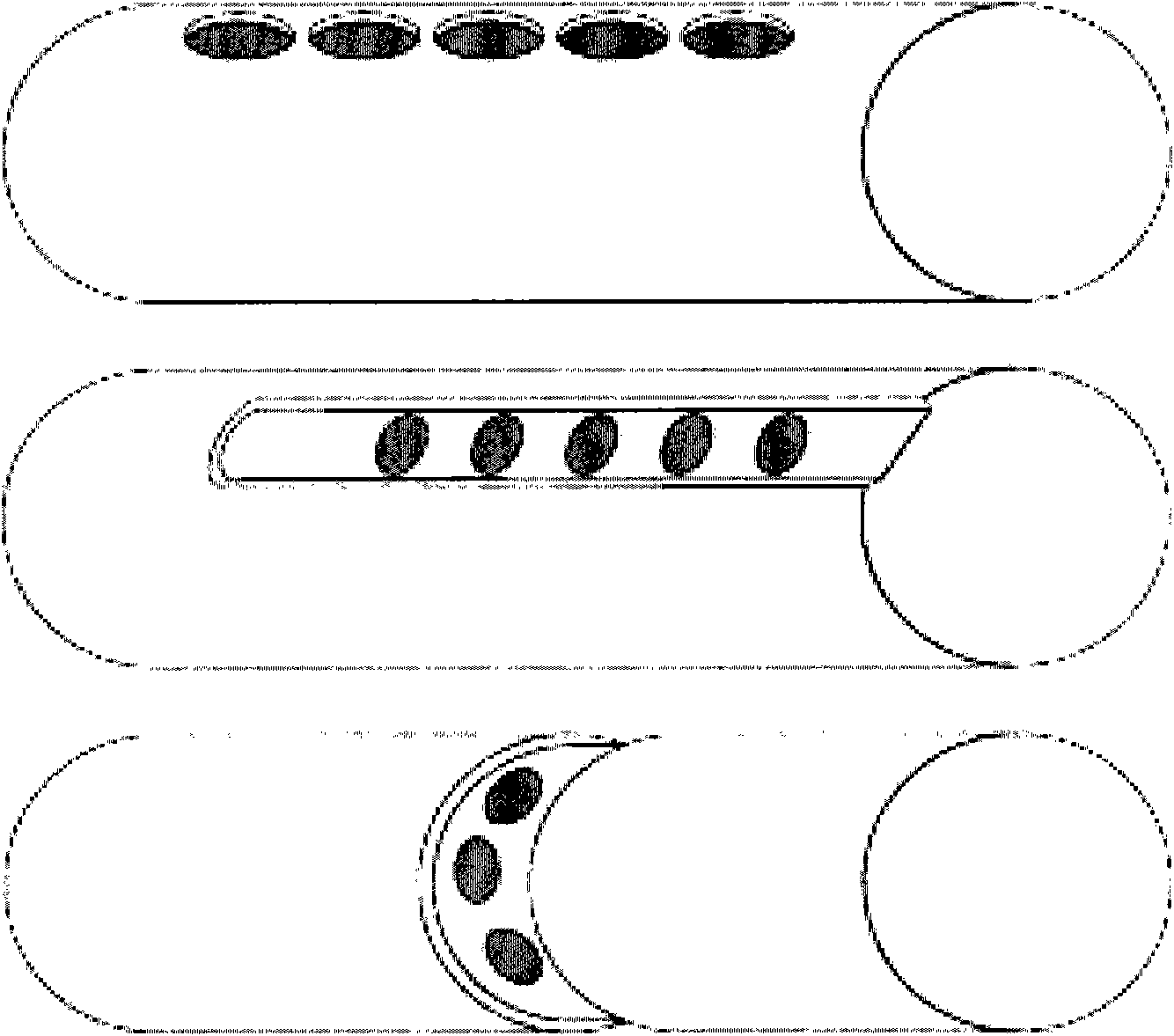
[0114] The surface of a nitinol-based metal guide (Euroflex) is structured to have specific sites, such as pores, where reactive groups are placed and where biochemical reactions occur ( figure 1 );

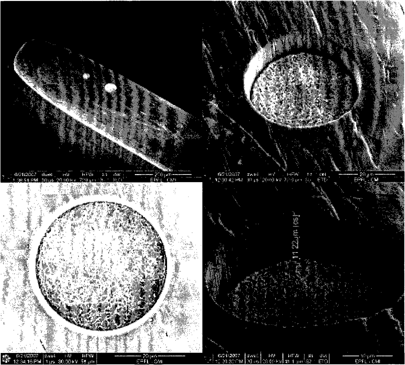
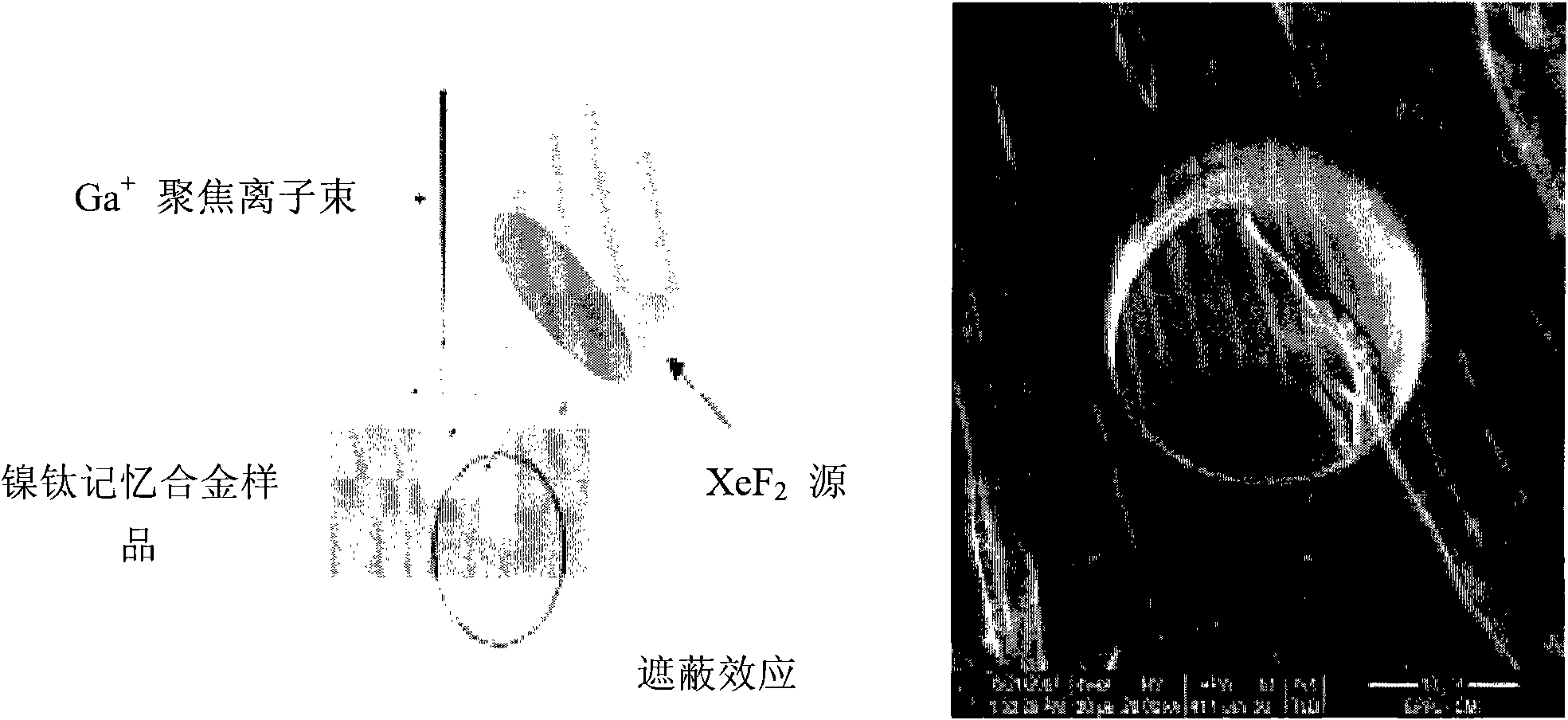
[0115] The "holes" can be obtained using various methods, such as focused ion beam lithography (FIB, Xie et al., Nuclear Instruments & Methods in Physics research Section B-beam Interactions with Materials and Atoms, 211(3): 363-368, 2003 ), by laser lithography followed by electrochemical etching and laser ablation.

[0116] Using FIB technology, a machine produces a beam of ions that is focused onto the surface that must be textured. Under the mechanical action of the ion beam, atoms of the surface material are removed from the surface. At the right time, the 8μm with a beam current of 20nA can be passed by FIB technology 3 the s -1 A hole with a diameter of 20 μm was form...

Embodiment 2

[0121] Example 2: Trauma caused by continuous in vivo insertion of metal guides into specific organs

[0122] A metal microguide (MTI 0.012" Silver speed) was used in these experiments and inserted into a microcatheter.

[0123] The device was introduced into a pig under general anesthesia at the suction level, followed by an intravascular route (via the femoral artery) up to the kidney at the level of the femoral triangle (scarpa). The guide is manipulated by follow-up observation of the device located in the femoral artery by arteriography.

[0124] When the device is at the entrance of the kidney, it can reach the kidney through the invading arterial route. This penetrates a few millimeters deep into the tissue and the device stays there for about 10 minutes.

[0125] Finally, remove the device.

[0126] After puncturing with the device according to the invention, the animals were sacrificed and their kidneys were taken as samples to evaluate their condition.

[01...

Embodiment 3

[0129] Example 3: Minimal invasiveness associated with device insertion in the liver

[0130] Unlike Example 1, the metal microguide used (MTI 0,012"Silver speed) was set in the fiberscope.

[0131] The device was introduced into a pig under general anesthesia at the suction level, followed by an intravascular route (via the femoral artery) up to the liver at the level of the femoral triangle. The guide is manipulated angiographically by said device located in the femoral artery.

[0132] When approaching the liver, the device is introduced into the organ. The puncture is a few millimeters deep into the tissue, and the device remains there again for about 10 minutes.

[0133] Finally, remove the device.

[0134] As previously mentioned, liver samples from this procedure can be used to assess the degree of involvement of this organ by the procedure.

[0135] There was no visible damage to the liver surface. When cut, two hemorrhagic nodes of 1.5 × 0.4 cm and 1.8 × 0.5 cm i...


PUM


Abstract

The device has a microinvasive or micro-sampling investigating system comprising a flexible metallic guide with an end containing a series of pits. The pits are directly coupled to a specific reactive group of a substrate, where the end is perforated. Another end of the metallic guide is used to maneuver the metallic guide and associated with an aspiration system. A removable protection system e.g. flexible catheter, is arranged at the former end of the metallic guide. An independent claim is also included for a method for detecting ex vivo of a substrate present in a tissue or organ.

Description

technical field [0001] The invention relates to the functioning of a transbody wall examination device, a method for analyzing substrates in vitro using the functionalized device of the invention, and the use of such a device in the manufacture of a device for diagnosing cancer, infection, inflammation in a patient , neurodegenerative disease or the use of a tool for transplant rejection. Background technique [0002] Examination or in vivo treatment devices are known in the art. Such devices are in the form of rigid tubes of the endoscopic type or catheters made of flexible tubes, which are inserted into the living body, in particular via natural pathways or blood vessels, and make them reach organs or specific tissues. These devices can specifically eliminate blood clots, or when they are combined with optical fibers, they can visualize and control the state of a system (such as the digestive tract) or an organ (such as the colon) in vivo. Then, the trauma to the patient...

Claims


Application Information

Patent Timeline
Patent Type & Authority Patents(China)
IPC IPC(8): A61B5/00
CPCA61B5/00A61B2562/043A61B5/6851A61B2562/02A61B5/1473A61B5/14542A61B5/4076
Inventor 阿兰·蓬皮杜阿尔贝-克洛德·贝纳姆欧
Owner BIOSTEMS LTD
Why Eureka
Try Eureka