(Classical swine fever virus) CSFV E2 protein ligand epitope peptide and application thereof
A swine fever virus, epitope peptide technology, applied in the field of molecular pathology and immunology, can solve the problem of no detailed report and so on
- Summary
- Abstract
- Description
- Claims
- Application Information
AI Technical Summary
Problems solved by technology
Method used
Image
Examples
Embodiment 1
[0064] Example 1, Design and synthesis of CSFVE2 protein polypeptide and Sulfo-NHS-Biotin labeled polypeptide
[0065] 1.1 Design of peptides
[0066] The amino acid sequence of the CSFVE2 protein was analyzed using DNASIS (Ver.2.5) software, and a polypeptide was designed, which contained part of the amino acid sequence of the E2 protein, and Cys was added to the N-terminus of the polypeptide, so as to be coupled to the carrier with the SMCC bifunctional reagent. Use the peptide program to analyze the difficulty of synthesis; design the synthesis (cleavage) program; edit the peptide chain; calculate and prepare solutions and reagents; start the synthesis program.
[0067] 1.2 Synthesis of peptides
[0068] Peptides were synthesized by solid-phase peptide synthesis on Fmoc-Amino Acids attached to Wang Resin (Fmoc-Amino Acids attached to Wang Resin, Shanghai Jier) using a Symphony 12-channel peptide synthesizer, and the peptides were synthesized according to standard operating...
Embodiment 2
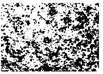
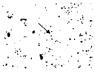
[0075] Example 2, Cell Immunochemical Staining Identification Biotin-SE24 Polypeptide Binding Test to PK-15 Cells
[0076] (1) Culture PK-15 cells at 37°C, 5% CO 2 After culturing in the incubator for 24 hours, wait for the cells to cover the 96-well cell culture plate;
[0077] (2) Pour off the culture supernatant, wash the cultured monolayer cells three times with pre-cooled PBS, and add concentrations of 0.2mmol / L, 0.1mmol / L, 0.05mmol / L and 0.025mmol / L to the culture plate respectively. L of Biotin-labeled polypeptide Biotin-SE24, 100 μL / well, do 3 replicates for each dilution, incubate at 4°C for 1 hour to fully combine the Biotin-labeled polypeptide with PK-15 cells, set 6 wells without adding Biotin-labeled polypeptide Normal cells were used as a control, and Biotin-labeled BSA was used as an irrelevant polypeptide control;
[0078] (3) Gently wash the cells 3 times with pre-cooled PBS to wash away unbound peptides and stop the peptide binding test;
[0079] (4) Fixed...
Embodiment 3
[0084] Example 3, Biotin-SE24 polypeptide binding assay and virus blocking assay analyzed by flow cytometry
[0085] The following experiments were performed simultaneously with PK-15 cells. The test is divided into 3 groups, test group A: Biotin-labeled polypeptide binding PK-15 cell test; test group B: Biotin-BSA binding PK-15 cell test, as a negative control; test group C: CSFV blocking PK-15 cell binding site After spotting, the Biotin-labeled polypeptide binding PK-15 cell test was carried out as a virus blocking test. The specific test steps are as follows:
[0086] (1) Culture PK-15 cells, after 24 hours of culture, the cells grow into a monolayer;
[0087] (2) Wash the cells 3 times with pre-cooled PBS, digest the cells with trypsin for about 20 minutes, blow the cells from the cell flask with a 10ml pipette, mix well to reduce the large cell clumps, centrifuge at 1500r / m for 5 minutes, discard decellularized supernatant;
[0088] (3) Add new pre-cooled PBS and res...
PUM
| Property | Measurement | Unit |
|---|---|---|
| fluorescence | aaaaa | aaaaa |
| fluorescence | aaaaa | aaaaa |
| fluorescence | aaaaa | aaaaa |
Abstract
Description
Claims
Application Information
 - R&D
- Intellectual Property
- Life Sciences
- Materials
- Tech Scout
- Unparalleled Data Quality
- Higher Quality Content
- 60% Fewer Hallucinations
Browse by: Latest US Patents, China's latest patents, Technical Efficacy Thesaurus, Application Domain, Technology Topic, Popular Technical Reports.
© 2025 PatSnap. All rights reserved.Legal|Privacy policy|Modern Slavery Act Transparency Statement|Sitemap|About US| Contact US: help@patsnap.com


