Direct PCR (Polymerase Chain Reaction) method without extracting animal tissue DNA (Deoxyribonucleic Acid)

A technology of animal tissue and tissue, applied in the field of PCR amplification, to achieve the effect of rapid cost, cost saving and simple method

Inactive Publication Date: 2012-02-08
CHINA AGRI UNIV
View PDF2 Cites 27 Cited by
  • Summary
  • Abstract
  • Description
  • Claims
  • Application Information

AI Technical Summary

Problems solved by technology

However, it is not feasible to use whole animal tissues such as ear tissues and hair follicles directly as templates for PCR amplification

Method used


Image

  • Direct PCR (Polymerase Chain Reaction) method without extracting animal tissue DNA (Deoxyribonucleic Acid)
  • Direct PCR (Polymerase Chain Reaction) method without extracting animal tissue DNA (Deoxyribonucleic Acid)
  • Direct PCR (Polymerase Chain Reaction) method without extracting animal tissue DNA (Deoxyribonucleic Acid)

Examples


Embodiment 1

[0021] Configure four different tissue lysates:

[0022] Lysis buffer 1: 10mM Tris-Cl pH8.0, 1mM EDTA pH8.0, 200μg / ml proteinase K;

[0023] Lysis solution 2: 10mM Tris-Cl pH8.0, 1mM EDTA pH8.0, 200μg / ml proteinase K, 0.1% SDS;

[0024] Lysis buffer 3: 10mM Tris-Cl pH8.0, 1mM EDTA pH8.0, 200μg / ml proteinase K, 0.01M NaCl;

[0025] Lysis solution 4: 10mM Tris-Cl pH8.0, 1mM EDTA pH8.0, 200μg / ml proteinase K, 0.1% SDS, 0.01M NaCl;

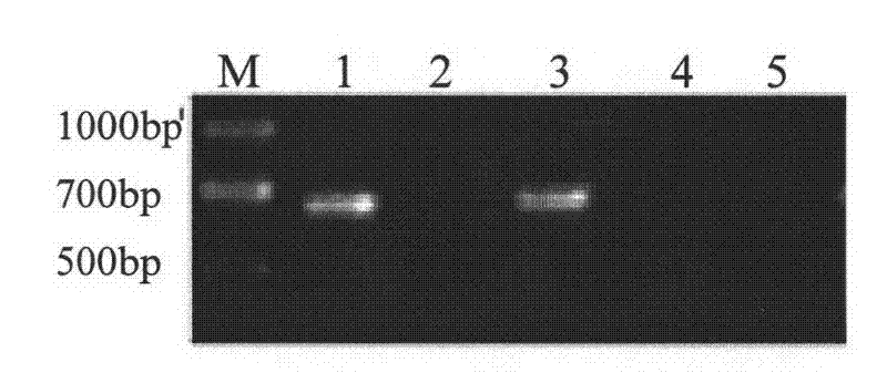
[0026] Take 0.01g of ear tissue, put it into a clean 200μl PCR tube, shred it fully, then take 50μl of the above four different lysates, first incubate at 55°C for 30min, and then denature at 95°C for 5min to inactivate proteinase K. Finally, 2 μl of four different digestion solutions were directly taken as templates, and the ear tissue without any lysate treatment was used as a control for PCR amplification to amplify the porcine myhc IIb gene fragment.

[0027] PCR reaction system: final concentration of 250μM dNTP, 10mM Tris-Cl, pH8.3, 50mM KCl,...

Embodiment 2

[0031] Lysis buffer: 10mM Tris-Cl pH8.0, 1mM EDTA pH8.0, 100μg / ml proteinase K, 0.01M NaCl

[0032] Take 0.01g of ear tissue, put it into a clean 200μl PCR tube, shred it fully, then take 50μl of the above-mentioned lysate, incubate at 37°C for 25min, then denature at 95°C for 5min to inactivate proteinase K, and finally directly Take 2 μl of digestion solution as a template, and use ear tissue without any lysate treatment as a control for PCR amplification to amplify the porcine myhc IIb gene fragment. The PCR reaction system and reaction conditions are the same as in Example 1.

[0033] After the amplification, 5 μl of the amplification product was taken for agarose gel analysis. It was found that a fragment of about 700 bp could be amplified from the ear tissue digested with the lysate, but no band could be amplified from the undigested ear tissue.

Embodiment 3

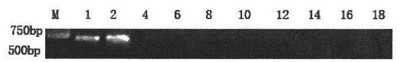
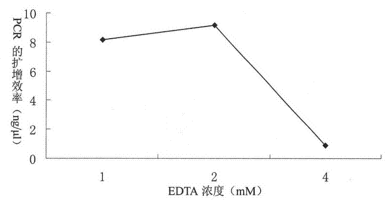
[0035] Optimize the concentration of EDTA in lysate 1: 10mM Tris-Cl, pH8.0, 1mM EDTA, pH8.0, 200μg / ml proteinase K, settings 1, 2, 4, 6, 8, 10, 12, 14, 11 different concentrations of 16, 18, 20mM. The treatment method of the ear tissue and the PCR reaction system and conditions are the same as in Example 1. Three treatments were performed for each concentration, and three repeated PCR amplifications were performed for each treatment to amplify the porcine myhc IIb gene fragment. After the PCR product was detected by agarose gel, it was found that no band could be detected after the concentration of 6mM ( figure 2 ). The PCR products of the first three concentrations were purified by agarose gel electrophoresis, and the purified products were determined by NanoDrop 2000 (NanoDrop Technologies, Wilmington, DE, USA). The results show that 2mM EDTA is the optimal concentration ( image 3 ).


PUM


Abstract

The invention relates to a direct PCR (Polymerase Chain Reaction) method without extracting animal tissue DNA (Deoxyribonucleic Acid). The method comprises the following steps of: digesting animal tissues with tissue lysis solution; and directly performing PCR amplification by using digestion solution as a template, wherein the used tissue lysis solution consists of Tris-C1, EDTA (Ethylene Diamine Tetraacetic Acid), protease K and NaCl. Compared with the conventional common method, the method has the advantages that: the method can be used for simultaneously treating a large number of animal tissue samples; high-flux molecular operation based on PCR amplification is realized; operation steps are quickly simplified; and cost is saved.

Description

technical field [0001] The present invention relates to the field of animal molecular biology, in particular to rapid digestion of animal tissues including ear tissue, blood or hair by using a specific tissue lysate, and then directly using a small amount of the digestion solution as a template to realize PCR amplification. Background technique [0002] The preparation of genomic DNA is essential to complete PCR amplification. In animal genetics and breeding, PCR amplification is the basis for research work such as gene polymorphism analysis, microsatellite analysis, gene cloning and sequencing. Currently, animal tissues commonly used for DNA extraction include ear tissue, blood and hair. Usually, the traditional phenol-chloroform extraction method is used to extract DNA from animal tissue samples, but this method has disadvantages such as cumbersome steps, time-consuming, reagent-consuming and poor safety. If a large number of animal tissue samples are extracted with this ...

Claims


Application Information

Patent Timeline
Patent Type & Authority Applications(China)
IPC IPC(8): C12N15/10C12Q1/68
Inventor 吴常信李红伟徐海岳赵春江
Owner CHINA AGRI UNIV
Why Eureka
Try Eureka