

Thromboprotein 1, thrombin 2, interleukin 17b receptor and heparin-binding epidermal growth factor-like growth factor associated with stem cell activity and their uses
A technology for coagulation of thrombus proteins and stem cells, which can be used in cell culture active agents, animal cells, vertebrate cells, etc., and can solve problems such as unproven stem cells
- Summary
- Abstract
- Description
- Claims
- Application Information
AI Technical Summary
Problems solved by technology
Method used
Image

Examples
example 1
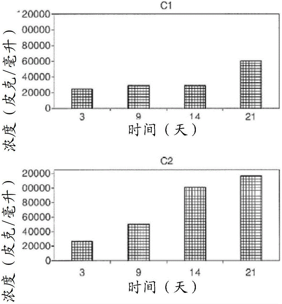
[0105] Example 1: Identification of secreted proteins specifically induced in UCB-MSCs by synovial fluid of arthritic patients
[0106] To identify substances that regulate cartilage regeneration and cartilage inflammation produced by UCB-MSCs, joint fluid from arthritic patients was added to the medium in which UCB-MSCs were cultured to a final concentration of 20% (v / v), followed by further culturing the resulting Product 3 hours. The obtained culture supernatant was used as an analysis sample. In addition, a UCB-MSC culture cultured without adding joint fluid and / or a medium containing 20% (v / v) joint fluid in which UCB-MSC was not cultured was used as a control group. Obtaining joint fluid from degenerative arthritis patients.
[0107] Proteins expected to be contained in each obtained culture or control sample are labeled with a detectable marker. The marker is biotin, and biotin is detected by fluorescent detection of a complex formed by specific binding between bio...
example 2
[0109] Example 2: Association of UCB-MSC Chondrogenic Differentiation with TSP-2
[0110] In this example, the association of UCB-MSC chondrogenic differentiation with TSP-2 was assessed. Additionally, it was assessed whether TSP-2 induced UCB-MSCs to differentiate into chondrocytes.
[0111] 1) Chondrogenic differentiation ability of various types of UCB-MSCs
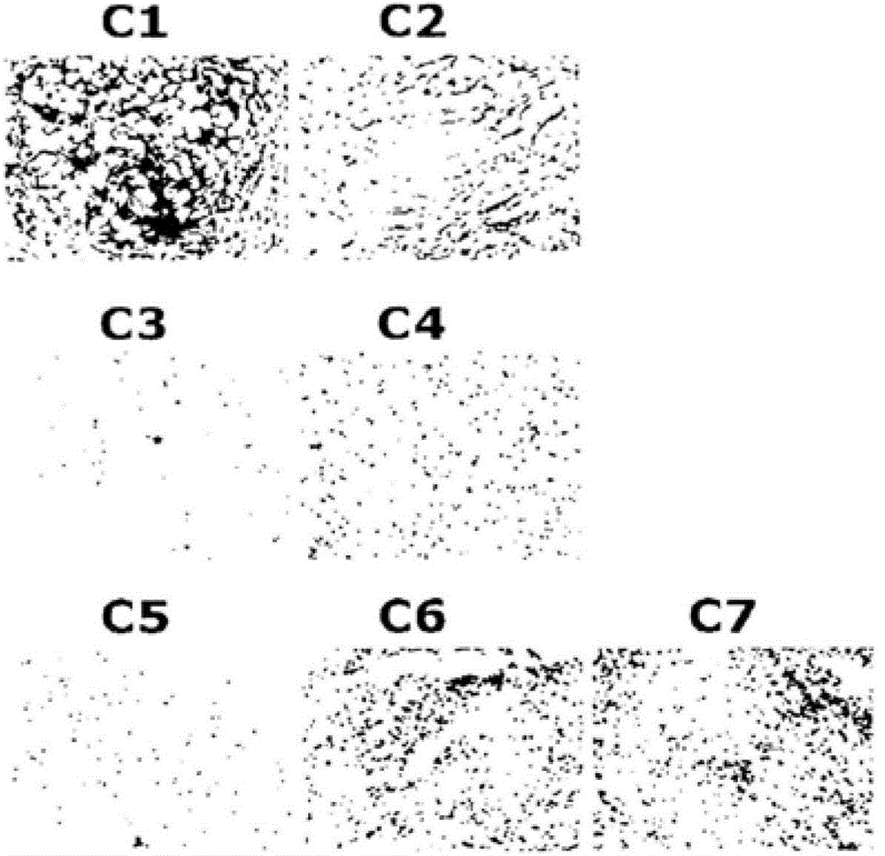
[0112] First, the chondrogenic differentiation ability of various types of UCB-MSCs was confirmed. Various types of UCB-MSCs were cultured in clumps in chondrogenic differentiation medium. Chondrogenic differentiation medium is containing 50 μg / ml ascorbate, 0.1 μM dexamethasone, 40 μg / ml L-proline, 100 μg / ml pyruvate, 10 ng / ml TGF-β3, 500 nM High glucose DMEM with g / ml BMP-6, 50 mg ITS+ / ml and 50 μg / ml gentamicin. The initial cell concentration was 5 x 10 per mL 5 cells and cultured in 15 ml polypropylene tubes for 4 weeks. The medium was changed twice a week and the pellets were fixed with 4% paraformaldehyde i...
example 3
[0122] Example 3: Expression level of TSP-2 according to chondrogenic differentiation ability
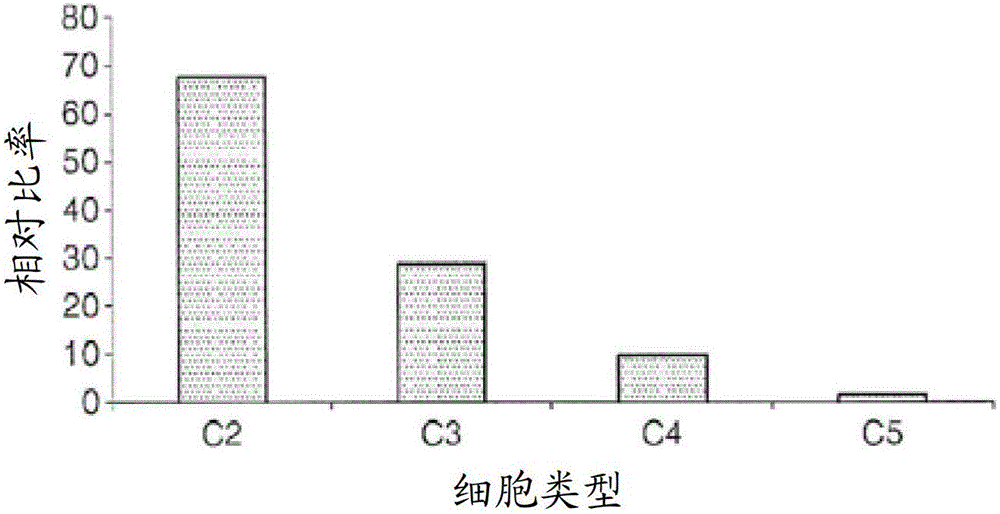
[0123] In this example, the expression level of TSP-2 of UCB-MSCs according to their chondrogenic differentiation ability was measured. First, C3, C4 and C5 UCB-MSCs were respectively cultured in chondrogenic differentiation medium under the same conditions for 7 days to induce chondrogenic differentiation. The relative chondrogenic differentiation capacity of C3, C4, and C5 UCB-MSCs has been experimentally confirmed previously and satisfies the condition of C3>C4>C5. Next, TSP-2 in the obtained culture supernatant was measured by ELISA.
[0124] Image 6 is a graph showing TSP-2 expressed in each of the culture supernatants of 3 types of UCB-MSCs cultured in a chondrogenic differentiation medium according to an example of the present invention. see Image 6 , C5UCB-MSCs classified as having the lowest chondrogenic differentiation capacity were secreted per 1 × 10 5 72 pg / ml TS...
PUM
| Property | Measurement | Unit |
|---|---|---|
| molecular weight | aaaaa | aaaaa |
| molecular weight | aaaaa | aaaaa |
| molecular weight | aaaaa | aaaaa |
Abstract
Description
Claims
Application Information