Method for detecting activity of O6-methylguanine-DNA (Deoxyribose Necleic Acid) methyltransferase
A technology of methyltransferase and methylguanine, applied in measuring devices, instruments, scientific instruments, etc.
- Summary
- Abstract
- Description
- Claims
- Application Information
AI Technical Summary
Problems solved by technology
Method used
Image

Examples
Embodiment 1
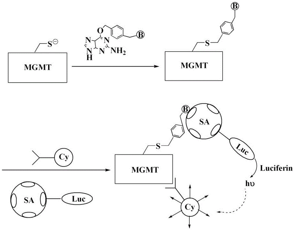
[0030] Example 1 O 6 -Methylguanine-DNA methyltransferase (MGMT) biotin labeling
[0031] In 4ml TBS buffer solution (pH 6.4) containing 4mg MGMT to be tested (taken from tumor cells) was added 1ml containing 2mg biotin-labeled O 6 -TBS buffer solution of benzylguanine (pH 6.4), mix well and place in a water bath at 37°C for 4 hours. After the reaction is over, add the reaction solution to a 14KD dialysis bag, and use a TBS buffer solution of pH 6.4 as the dialysate Dialysis was performed at 4°C, and the dialysate was changed every 3 hours, for a total of three changes, then the retentate was filtered with cross-linked agarose gel CL-6B, the filtrate was taken, and freeze-dried at 4°C for 10 hours to obtain biotin-labeled O 6 -Methylguanine-DNA methyltransferase.
Embodiment 2
[0032] Example 2 Detection of MGMT activity
[0033] (1) Coating: The streptavidin-firefly luciferase fusion protein was prepared with a 0.1mol / L TBS buffer solution with a pH of 6.4 to make 50ml of a 1mg / mL coating solution. Add the coating solution to the wells of a 96-well white-bottomed polystyrene microtiter plate, 200μl per well, place at 37°C for 60 minutes, discard the coating solution, and add 200μl of 10% bovine serum albumin (BSA) to each well. The TBS buffer solution of) was used as the blocking solution, sealed at 37°C for 1 hour, and the plate was washed with the above TBS buffer solution, soaked for 3 minutes each time, washed three times, dried under vacuum at 30°C, and stored in a closed bag under vacuum to prepare Microplate coated with streptavidin-firefly luciferase fusion protein;
[0034] (2) Add samples: add the streptavidin-firefly luciferase fusion protein-coated microtiter plate prepared in step (1) to the sample holes of the microtiter plate prepared in ...
Embodiment 3
[0039] Example 3 Detection of MGMT activity
[0040] (1) Coating: The streptavidin-alkaline phosphatase fusion protein was used to prepare 50 ml of a 3 mg / mL coating solution with a 0.5 mol / L TBS buffer of pH 6.4. Add the coating solution to the loading wells of a 96-well white-bottomed polystyrene microtiter plate, 200μl per well, and place at 25°C for 1h. Discard the coating solution, and add another 200μl mass concentration of 5% bovine serum albumin to each well ( BSA) the above TBS buffer as a blocking solution, blocked at 37°C for 1 hour, washed with the above TBS buffer, soaked for 3 minutes each time, washed the plate 3 times, dried under vacuum at 25°C, and stored in a closed bag under vacuum. Streptavidin-alkaline phosphatase fusion protein;
[0041] (2) Sample addition: Add the concentration of 1000, 500, 250, 125, 62.5, 62.5, 1000, 500, 250, 125, 62.5, to the sample well of the microtiter plate coated with streptavidin-alkaline phosphatase fusion protein prepared in st...
PUM
 Abstract
Description
Claims
Application Information
 - R&D
- Intellectual Property
- Life Sciences
- Materials
- Tech Scout
- Unparalleled Data Quality
- Higher Quality Content
- 60% Fewer Hallucinations
Browse by: Latest US Patents, China's latest patents, Technical Efficacy Thesaurus, Application Domain, Technology Topic, Popular Technical Reports.
© 2025 PatSnap. All rights reserved.Legal|Privacy policy|Modern Slavery Act Transparency Statement|Sitemap|About US| Contact US: help@patsnap.com