
Preparation method of micro/nanofiber sustained release preparation

A nanofiber and sustained-release preparation technology, applied in the field of biomedicine, can solve the problems that the electrospinning fiber film is difficult to achieve long-term drug release, the specific surface is too large, etc., and achieves the effect of long release time, less drug amount, and improved curative effect.

Inactive Publication Date: 2013-05-15
崔文国 +3
View PDF2 Cites 9 Cited by
  • Summary
  • Abstract
  • Description
  • Claims
  • Application Information

AI Technical Summary

Problems solved by technology

Unfortunately, it is difficult to achieve long-term drug release using electrospun fibrous films of existing biodegradable polymers because of their large surface-area-to-volume ratio.

Method used


Image

  • Preparation method of micro/nanofiber sustained release preparation
  • Preparation method of micro/nanofiber sustained release preparation
  • Preparation method of micro/nanofiber sustained release preparation

Examples


Embodiment 1

[0038] Weigh 2g of IBU and completely dissolve it in 100mL of n-hexane, weigh 4g of modified mesoporous silica nanoparticles (MMS) and add them to the IBU solution, incubate at 37°C for 24h, centrifuge, take the solid and dry to obtain MMS-IBU nanoparticles;

[0039] Disperse MMS-IBU nanoparticles in a mixed solvent of ethanol and hexafluoroisopropanol (HFIP), weigh 40g of poly-L-lactic acid (PLLA) and dissolve it in DCM, mix the two solutions, and control the PLLA content to 22 wt%;

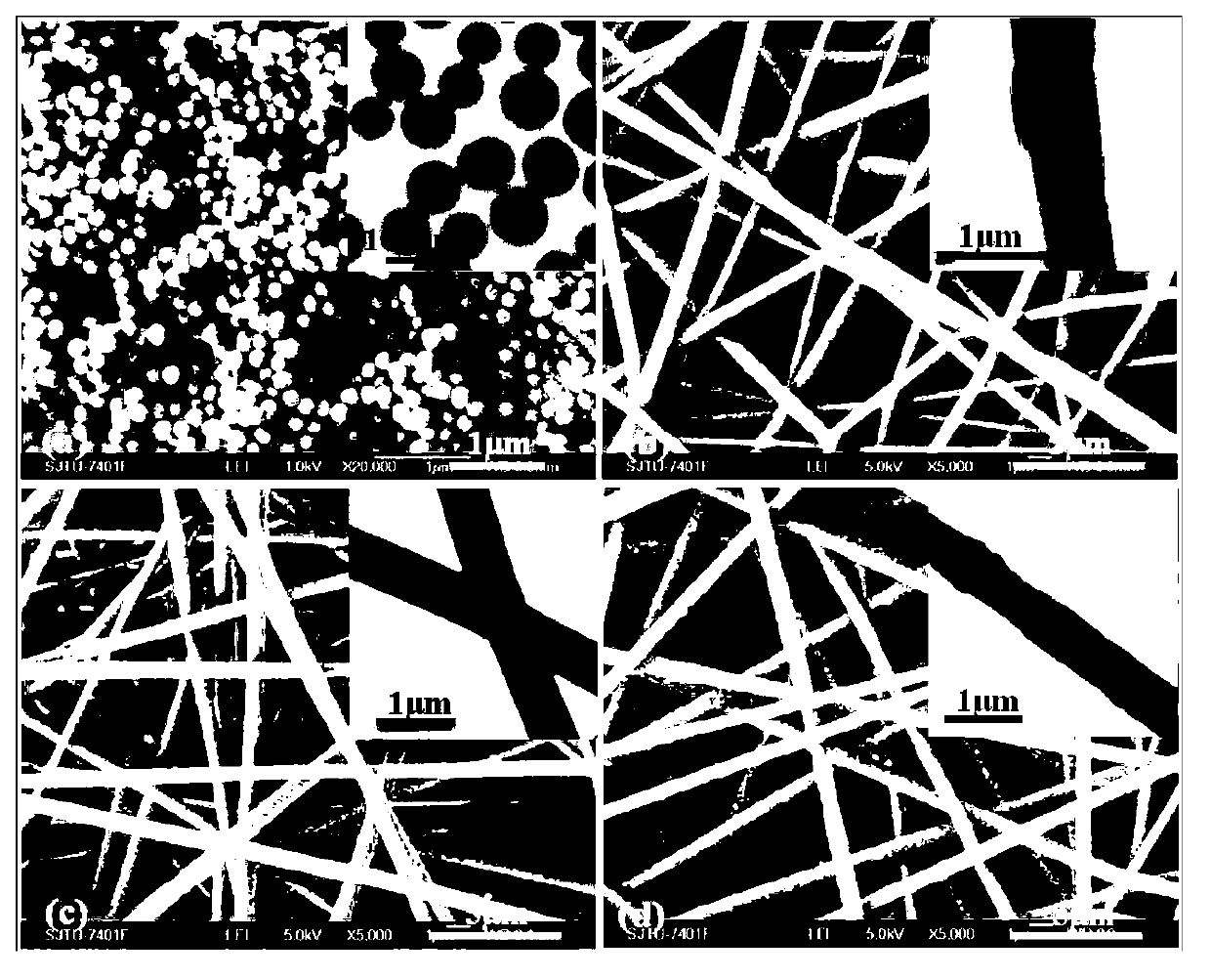
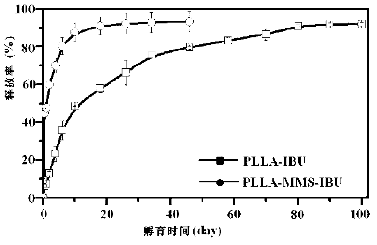
[0040] Electrospinning to obtain ibuprofen micro / nanofiber sustained-release preparation (PLLA-MMS-IBU). Among them, the electrospinning process parameter conditions are: voltage 11kV, flow rate 0.04mL / min, and the distance between the receiving table and the spinneret is 15cm.

[0041] PLLA-IBU (with an IBU content of 9 wt%) and electrospun PLLA were prepared by the same preparation process.

[0042] Scanning electron microscope (SEM) and transmission electron microscope (TEM) observed the obt...

Embodiment 2

[0047] Weigh 2g of IBU completely dissolved in 100mL of n-hexane, weigh 3g of modified mesoporous silica nanoparticles (MMS) into the IBU solution, incubate at 37°C for 24h, centrifuge, take the solid and dry to obtain MMS-IBU nanoparticles;

[0048] Disperse MMS-IBU nanoparticles in a mixed solvent of ethanol and HFIP, weigh 38g poly-L-lactic acid (PLLA) and dissolve it in DCM, mix the two solutions, and control the PLLA content to 22 wt%;

[0049] Electrospinning to obtain ibuprofen micro / nanofiber sustained-release preparation (PLLA-MMS-IBU). Among them, the electrospinning process parameter conditions are: voltage 11kV, flow rate 0.04mL / min, and the distance between the receiving table and the spinneret is 15cm.

Embodiment 3

[0051] Weigh 2.2g of IBU and completely dissolve it in 100mL of n-hexane, weigh 4g of modified mesoporous silica nanoparticles (MMS) and add them to the IBU solution, incubate at 37°C for 24h, centrifuge, take the solid and dry to obtain MMS-IBU nanoparticles;

[0052] Disperse the MMS-IBU nanoparticles in the mixed solvent of ethanol and HFIP, weigh 40g poly-L-lactic acid (PLLA) and dissolve it in DCM, mix the two solutions, and control the PLLA content to 22 wt%;

[0053] Electrospinning to obtain ibuprofen micro / nanofiber sustained-release preparation (PLLA-MMS-IBU). Among them, the electrospinning process parameter conditions are: voltage 11kV, flow rate 0.04mL / min, and the distance between the receiving table and the spinneret is 15cm.


PUM

PropertyMeasurementUnit
molecular weightaaaaaaaaaa

Abstract

The invention provides a preparation method of a micro / nanofiber sustained release preparation. The preparation method of the micro / nanofiber sustained release preparation comprises the following steps of: dissolving medicaments in an organic solvent, then adding modified mesoporous silica nano particles, and incubating; separating solids; dispersing the solids into the organic solvent, dissolving degradable high molecule polymers into the organic solvent, and mixing the two solutions; and spinning the mixed solution by an electrostatic spinning method, thereby obtaining the micro / nanofiber sustained release preparation. The micro / nanofiber sustained release preparation prepared through the preparation method can effectively control the slow release of medicaments, and the degradable high molecule polymers can degrade automatically; the preparation is non-toxic, has no immunogenicity, has good biocompatibility, improves the curative effect, and reduces the side effects; and different medicaments can be packaged by utilizing the preparation method, and meanwhile fiber supports for packaging different medicaments can be obtained; the long-time release of the medicaments has important significance clinically, and the method possibly become an ideal technical platform for controlling and releasing the medicament for a long time.

Description

technical field [0001] The invention belongs to the field of biomedicine, and in particular relates to a preparation method of a micro / nano fiber sustained-release preparation. Background technique [0002] Adhesion is the process of human tissue regeneration. When human tissue is cut open, interstitial fluid seeps out from the incision, and there is a large amount of tissue fibrin in it. They interweave each other to form a fibrous network, which penetrates with capillaries. Capillaries supply the newly formed tissue with nutrients, so that the new tissue structure continues to extend until the tissue is completely repaired. If a fibrin network is formed between abnormal tissues, fibrous adhesions are formed, causing dysfunction, which is a pathological process that should not exist. Almost all surgeries involve the problem of adhesions between tissues. Not only can it cause serious complications, but it is also one of the main reasons for significantly higher complicatio...

Claims


Application Information

Patent Timeline
Patent Type & Authority Applications(China)
IPC IPC(8): A61K9/14A61K47/04A61K45/00A61K31/192A61K33/38A61K47/34A61P31/04A61P29/00
Inventor 崔文国范存义刘珅胡昌敏
Owner 崔文国
Learn More