Cylinder support for repairing spinal cord injury and application method thereof
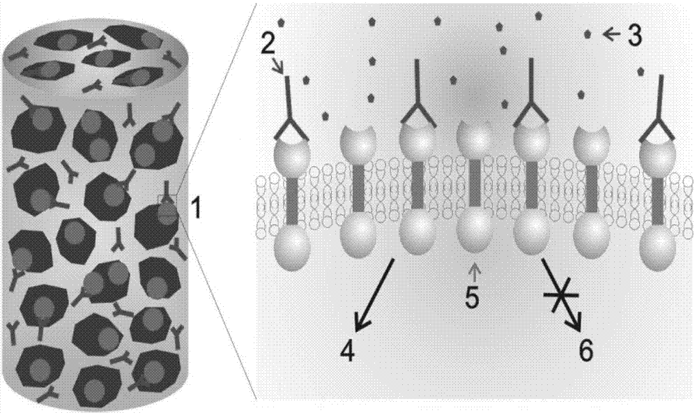
A spinal cord injury, cylinder technology, applied in the field of biomedical materials, can solve problems such as unsatisfactory clinical effects, achieve ultra-high porosity, promote growth, and promote spinal cord nerve regeneration.
- Summary
- Abstract
- Description
- Claims
- Application Information
AI Technical Summary
Problems solved by technology
Method used
Image
Examples
Embodiment 1
[0026] Embodiment 1-for the construction of the cylinder support of repairing spinal cord injury
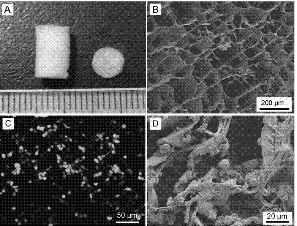
[0027] 1. Construction of Porous Collagen Sponge Cylindrical Scaffolds
[0028] Collagen raw materials were dissolved in 0.5mol / L acetic acid and allowed to stand at 4°C for 8 hours. After mixing evenly, 4mol / L NaOH was added for neutralization. The obtained homogeneous liquid was dialyzed with deionized water for 5 days, and the deionized water was changed every 3 hours during dialysis. ionized water. The dialyzed solution was put into a mold with a diameter of 4 mm, and freeze-dried to obtain a collagen sponge cylindrical scaffold.
[0029] 2. Construction of Porous Collagen Sponge Scaffolds Crosslinked with EGFR Antibody Cetuximab
[0030] Using Traut's Reagent (2-iminohydrothiol chloride) and Sulfo-SMCC (4-(N-maleimidomethyl)cyclohexane-1-carboxylic acid sulfosuccinimidyl ester sodium salt ) two-step coupling reaction to covalently cross-link cetuximab to the collagen scaf...
Embodiment 2
[0032] Example 2 - Characterization of Cylindrical Scaffold Materials for Repairing Spinal Cord Injuries
[0033] 1. Scanning electron microscope (SEM) to observe the morphology and pore size of the collagen scaffold
[0034] The collagen sponge scaffold was sprayed with gold in a dry state, observed and photographed under a scanning electron microscope. Observe the surface morphology, and take 5 fields of view in the material, and count the pore size and range.
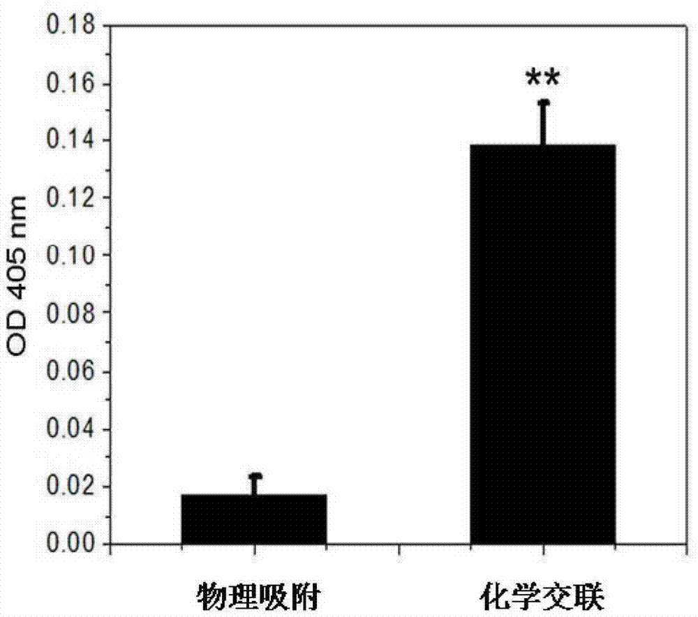
[0035] 2. Detection of Cetuximab loaded on the Col-cetuximab sponge scaffold
[0036] After cetuximab was covalently cross-linked or simply adsorbed to the collagen scaffold, the collagen scaffold was blocked with 5% BSA, then excess BSA was removed, washed three times with PBS, and alkaline phosphatase-labeled anti-mouse antibody was added, at 37°C Incubate for 1h. Aspirate the antibody, wash it three times with PBS, and then add 2 mg / mL p-nitrophenyl phosphate disodium salt (p-Npp) for color development, p-Npp i...
Embodiment 3
[0037] Example 3 - The in vitro induction and differentiation of NSCs by cylindrical scaffold materials used for repairing spinal cord injuries
[0038] 1. Extraction of Myelin Proteins
[0039] Prepared by discontinuous sucrose density gradient centrifugation. Spinal cords were obtained from adult rats, homogenized and crushed in 0.3mol / L sucrose solution, and then slowly spread on top of 0.85mol / L sucrose. The samples were centrifuged at 27,000 g for 1 h, and the sheath protein fragments were collected and extracted at the boundary of 0.32 / 0.85 mol / L sucrose. The samples were allowed to stand in deionized water for 1 h, and then collected by centrifugation at 12000 g. The obtained samples were filtered with a 0.22 μm filter membrane to remove large aggregates, sterilized, and stored at -80°C for use.
[0040] 2. Isolation and culture of primary NSCs and inoculation on scaffold materials
[0041] The telencephalon was isolated from SD suckling mice born at 12 hours, cut i...
PUM
| Property | Measurement | Unit |
|---|---|---|
| diameter | aaaaa | aaaaa |
| length | aaaaa | aaaaa |
| weight | aaaaa | aaaaa |
Abstract
Description
Claims
Application Information
 - R&D
- Intellectual Property
- Life Sciences
- Materials
- Tech Scout
- Unparalleled Data Quality
- Higher Quality Content
- 60% Fewer Hallucinations
Browse by: Latest US Patents, China's latest patents, Technical Efficacy Thesaurus, Application Domain, Technology Topic, Popular Technical Reports.
© 2025 PatSnap. All rights reserved.Legal|Privacy policy|Modern Slavery Act Transparency Statement|Sitemap|About US| Contact US: help@patsnap.com


