Dry-type immunoassay test strip and preparation method and application thereof

A technology for immune detection and test strips, applied in the field of clinical medical diagnosis, can solve the problems of complex process and long detection time, and achieve the effects of accurate detection results, shortened detection time and cost saving.

Inactive Publication Date: 2013-07-31
GETEIN BIOTECH
View PDF12 Cites 21 Cited by
  • Summary
  • Abstract
  • Description
  • Claims
  • Application Information

AI Technical Summary

Problems solved by technology

However, the Fusion5 membrane has a large pore size, and it is difficult to immobilize the antibody. It is necessary to prepare a special STONE to immobilize the antibody. The process is relatively complicated. At the same time, it takes ten minutes for the test strip chromatography prepared by the patent CN102192983A, and the detection time is long.

Method used


Image

  • Dry-type immunoassay test strip and preparation method and application thereof
  • Dry-type immunoassay test strip and preparation method and application thereof
  • Dry-type immunoassay test strip and preparation method and application thereof

Examples


Embodiment 1

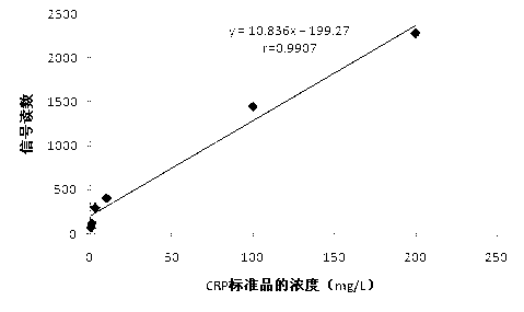
[0029] Example 1 Preparation and use of C-reactive protein colloidal gold detection test paper

[0030] 1. Preparation of C-reactive protein colloidal gold detection test paper

[0031] (1) Preparation of colloidal gold-antibody complex

[0032] a. Preparation of biotinylated C-reactive protein (CRP) monoclonal antibody complex: First, link 6-aminohexose to biotin to obtain long-arm biotin, add 80 μg of biotin per ml of mouse anti-human CRP monoclonal antibody Add and react for 1 hour to generate long-arm biotin N-hydroxysuccinamide ester, which is a biotinylated CRP monoclonal antibody complex, and dialyze through 1% BSA pH7.5 to remove free biotin;

[0033] b. Preparation of colloidal gold-labeled avidin: adjust the pH value of colloidal gold to 7.5 with 1% potassium carbonate solution, add streptavidin solution according to the amount of 10 μg streptavidin / ml colloidal gold, mix well, and place at 25°C After reacting in a water bath for 30 minutes, add 5% BSA, block for...

Embodiment 2

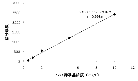
[0071] Example 2 Preparation and use of cystatin C fluorescence detection test strips

[0072] 1. Preparation of cystatin C (CysC) fluorescent test paper

[0073] (1) Cystatin C antibody labeled with fluorescent latex microspheres

[0074] a. Preparation of fluorescent latex microspheres: Dilute latex microspheres with a particle size of 400 nm to a final concentration of 30 mg / ml and a volume of 6 ml with an adsorption buffer (50 mmol / L, citrate buffer at pH 5.8) to obtain latex Microsphere suspension; add an appropriate amount of red fluorescein rhodamine-labeled streptavidin (purchased from Shanghai Enzyme Biotechnology Co., Ltd.) in the adsorption buffer, and the final volume is 6ml; add the above-mentioned latex microsphere suspension to Prepare a mixed solution in the above-mentioned adsorption buffer containing red fluorescein rhodamine-labeled streptavidin; incubate the resulting mixed solution at room temperature for 1-2 hours, and keep stirring, then centrifuge, c...

Embodiment 3

[0110] Example 3 Preparation and use of alpha-fetoprotein (AFP) fluorescent test strips

[0111] 1. Preparation of fluorescent test paper for alpha-fetoprotein detection

[0112] (1) Fluorescent latex microspheres labeled alpha-fetoprotein (AFP) antibody

[0113] a. Preparation of fluorescent latex microspheres: Dilute latex microspheres with a particle size of 200 nm to a final concentration of 30 mg / ml and a volume of 6 ml with an adsorption buffer (50 mmol / L, citrate buffer at pH 5.8) to obtain latex Microsphere suspension; add an appropriate amount of fluorescein Cy5 in the adsorption buffer, and the final volume is 6ml; add the above-mentioned latex microsphere suspension to the above-mentioned adsorption buffer containing fluorescein Cy5-labeled streptavidin to obtain Mixed solution: Incubate the resulting mixed solution at room temperature for 1-2 hours with constant stirring, then centrifuge to collect the precipitate, dissolve the precipitate in storage buffer (ads...


PUM


Abstract

The present invention relates to a dry-type immunoassay test strip and a preparation method and application thereof. On a plastic substrate of the test strip is sequentially stuck with a sample pad, a nitrocellulose membrane anda water absorption pad from left to right, wherein the right end of nitrocellulose membrane is provided with a control line and a detection line that are parallel to each other; the left end of nitrocellulose membrane is provided with a tracer particle labeled antibody/ antigen-coated line and a closed protein line I, wherein the closed protein line I is formed by coating closed protein solution I with a nitrocellulose membrane, and the antibody/ antigen-coated line is formed by spraying the tracer particle labeled antibody/ antigen on a closed protein line II, and the closed protein line II is formed by coating closed protein solution II with a nitrocellulose membrane. . The test strip can be applied in the immunoassay based on the double antibody sandwich method or the competitive method, and has advantages of simple operation, rapid chromatography, high specificity, accurate test results and strong stability.

Description

Technical field [0001] The invention is the field of clinical medical diagnosis, and it involves a dry immunohistic detection test note and its preparation methods and applications. Background technique [0002] Nowadays, dry immunoaged quantitative testing is generally tested by in vitro immune layer analysis reagents. The test note mainly combines the combination of affinable technology, imprinting technology, immunohistan label and layer analysis technology. Generally, it includes sample pads, blood filtration film, combinationThe four -to -five layers of pads, nitrate cellulose, and water absorption pads (four layers generally combine blood filtering function with sample pads), and the antibody packs of trace particles (colloidal gold, fluorescent, etc.) are combined with pads. [0003] The patented CN102192983A has changed the combination of traditional immunological analysis membranes and turned into a three -membrane system -Fusion5, nitrate cellulose film, and water absor...

Claims


Application Information

Patent Timeline
Patent Type & Authority Applications(China)
IPC IPC(8): G01N33/543G01N33/532G01N33/558
Inventor 苏恩本王勇焦丽蓉
Owner GETEIN BIOTECH
Why Eureka
Try Eureka