Preparation method of caprolactone lactate copolymer/collagen/chitosan small-caliber intravascular stent

A collagen and vascular stent technology, which is applied in the field of preparation of lactic caprolactone copolymer/collagen/chitosan small-diameter vascular stents, can solve the problem of poor biocompatibility of polymer nanofibers and inability to reach vascular tissue Engineering scaffold requirements, poor mechanical properties of natural materials, etc., to achieve the effect of promoting three-dimensional ingrowth of cells, excellent biocompatibility, and good mechanical properties

Active Publication Date: 2013-08-28
DONGHUA UNIV
View PDF4 Cites 13 Cited by
  • Summary
  • Abstract
  • Description
  • Claims
  • Application Information

AI Technical Summary

Problems solved by technology

However, the mechanical properties of natural materials are poor, and the biocompatibility of polymer nanofibers is not good, and because of the nanoscale fibers, it is difficult for cells to penetrate into the interior of the scaffold, which cannot meet the requirements of vascular tissue engineering scaffolds.
At present, there is no report to combine the three materials of caprolactone lactate copolymer, collagen, and chitosan through an improved electrospinning method to prepare a small-diameter vascular stent that mimics ECM in structure and function.

Method used


Image

  • Preparation method of caprolactone lactate copolymer/collagen/chitosan small-caliber intravascular stent
  • Preparation method of caprolactone lactate copolymer/collagen/chitosan small-caliber intravascular stent
  • Preparation method of caprolactone lactate copolymer/collagen/chitosan small-caliber intravascular stent

Examples


Embodiment 1

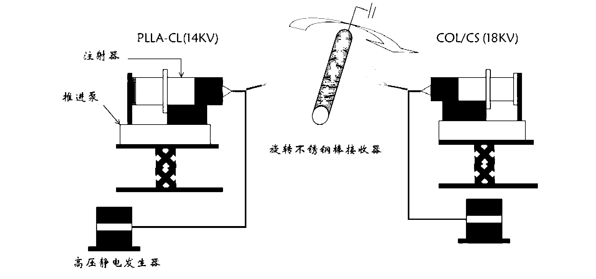
[0029] Take the caprolactone lactate copolymer and collagen and the chitosan of 0.3g with an electronic balance; the caprolactone lactone copolymer of 0.4g is dissolved in 5ml hexafluoroisopropanol, magnetically stirred until completely dissolved; dissolve 0.4g of collagen in 5ml hexafluoroisopropanol, and magnetically stir until completely dissolved; dissolve 0.3 of chitosan in 5ml hexafluoroisopropanol / trifluoroacetic acid (V / V, 9 / 1) Mix the solvents and stir until they are completely dissolved; after the three solutions are completely dissolved, the caprolactone lactone copolymer spinning solution is used as component 1, and 4.5mL of collagen spinning solution and 0.5mL of chitosan are used for spinning The solution is mixed evenly as component 2, and the two-way electrospinning method is used to suck the spinning solution of component 1 and component 2 into a 5mL syringe respectively, connect the No. The pump adjusts the flow rate (mL / h) of component 1 and component 2 as ...

Embodiment 2

[0031] Take the caprolactone lactate copolymer and collagen and the chitosan of 0.3g with an electronic balance; the caprolactone lactone copolymer of 0.4g is dissolved in 5ml hexafluoroisopropanol, magnetically stirred until completely dissolved; 0.4g of collagen was dissolved in 5ml hexafluoroisopropanol, magnetically stirred until completely dissolved; 0.3g of chitosan was dissolved in 5ml hexafluoroisopropanol / trifluoroacetic acid (V / V, 9 / 1) Mix the solvents and stir until they are completely dissolved; after the three solutions are completely dissolved, the caprolactone lactone copolymer spinning solution is used as component 1, and 4.5mL collagen spinning solution and 0.5mL chitosan spinning solution are taken. The silk liquid is mixed evenly as component 2, and the two-way electrospinning method is used to suck the spinning solution of component 1 and component 2 into a 5mL syringe respectively, connect the No. The syringe pump adjusts the flow rate (mL / h) of component ...


PUM

PropertyMeasurementUnit
diameteraaaaaaaaaa

Abstract

The invention relates to a preparation method of a caprolactone lactate copolymer/collagen/chitosan small-caliber intravascular stent, and the preparation method comprises the following steps of: dissolving a caprolactone lactate copolymer into a solvent to obtain a caprolactone lactate copolymer spinning solution; dissolving collagen into the solvent to obtain a collagen spinning solution; dissolving chitosan into the solvent to obtain a chitosan spinning solution; and carrying out bidirectional electrostatic spinning by taking the caprolactone lactate copolymer spinning solution as a component I and a mixed solution of the collagen spinning solution and the chitosan spinning solution which have the volume ratio of 5-10:1 as a component II to obtain the caprolactone lactate copolymer/collagen/chitosan small-caliber intravascular stent. The biomaterial designed into a multicomponent gradient symmetrical structure has the advantages of better mechanical property and excellent biocompatibility and especially has the advantages of potential on accelerating cell three-dimensional ingrowth and important application in small-caliber vascular tissue engineering.

Description

technical field [0001] The invention belongs to the field of preparation of tissue engineering small-diameter vascular stents, in particular to a preparation method of lactate caprolactone copolymer / collagen / chitosan small-diameter vascular stents. Background technique [0002] Electrospinning is a common technique for preparing fibers with diameters ranging from nanometer to micrometer, and nanostructured materials offer a great potential for tissue engineering. Because the diameter of nanoscale fibers is similar to the physical structure of protein fibers in natural extracellular matrix (ECM), and the nanofiber structure has high specific surface area and porosity, it can simulate the structure and function of ECM, which is conducive to cell adhesion, proliferation and tissue regeneration. In recent years, electrospinning technology has been applied to a variety of tissue engineering fields, such as skin, nerves, blood vessels, bone and cartilage, etc., especially in the ...

Claims


Application Information

Patent Timeline
Patent Type & Authority Applications(China)
IPC IPC(8): D04H1/4382D04H1/728A61L27/40
Inventor 莫秀梅吴桐黄晨
Owner DONGHUA UNIV
Why Eureka
Try Eureka