
3D printing artificial blood vessel and preparation method thereof

An artificial blood vessel, 3D printing technology, applied in the direction of blood vessels, human tubular structure devices, non-woven fabrics, etc., can solve the problems of small diameter of the tube wall, easy to seep blood, and difficult for cells to grow in three-dimensionally, and achieves simple and economical operation. High efficiency and good repeatability

Pending Publication Date: 2020-04-14
SHANGHAI CHANGDY MEDICAL CO LTD
View PDF4 Cites 1 Cited by
  • Summary
  • Abstract
  • Description
  • Claims
  • Application Information

AI Technical Summary

Problems solved by technology

[0003] Currently commercialized artificial blood vessels still have various problems: the artificial blood vessels made of expanded polytetrafluoroethylene (ePTFE) have needle holes in the sutures, which are prone to bleeding during and after the operation; and the diameter of the tube wall is relatively small. Small, cells are not easy to grow into three-dimensionally
Moreover, the above-mentioned commercial artificial blood vessels are all standardized products, and the diameter and wall thickness of the blood vessels are fixed, so it is difficult to fully match the actual diseased blood vessel size of each patient.
[0004] At present, there is no artificial blood vessel that can meet the requirements of no blood leakage during the operation, bionic extracellular matrix of the blood vessel wall, conducive to the three-dimensional growth of cells, and can be customized according to the actual size of the diseased blood vessel of the patient.

Method used


Image

  • 3D printing artificial blood vessel and preparation method thereof
  • 3D printing artificial blood vessel and preparation method thereof
  • 3D printing artificial blood vessel and preparation method thereof

Examples


preparation example Construction

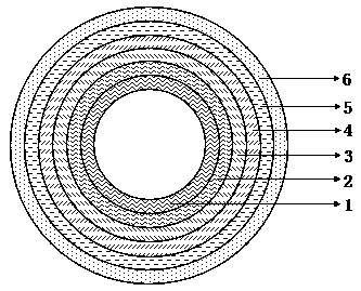
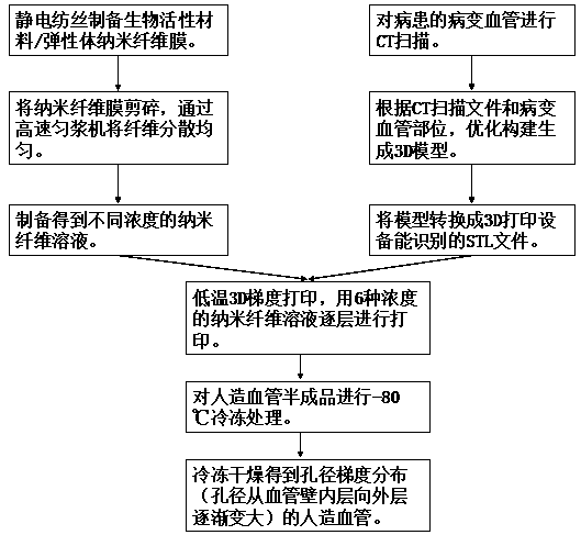
[0032] like figure 1 , the 3D printed artificial blood vessel contains a six-layer structure, which is the first layer, the second layer, the third layer, the fourth layer, the fifth layer, and the sixth layer from the inside to the outside. The preparation method is as follows: figure 2 ,include

[0033] 1) Prepare bioactive material / elastomer polymer composite nanofibrous membrane by electrospinning.

[0034] 2) Shredding the nanofiber membrane, placing it in a solvent and using a high-speed homogenizer to disperse the fibers evenly;

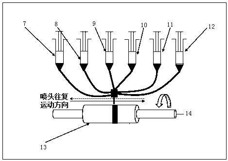
[0035] 3) Prepare nanofiber solutions for 3D printing with different concentrations, which are the first solution 7, the second solution 8, the third solution 9, the fourth solution 10, the fifth solution 11 and the sixth solution 12, such as image 3 .

[0036] 4) Use 3D printing technology to prepare semi-finished artificial blood vessels by printing nanofiber solutions with different concentrations layer by layer at low temperature;

...

Embodiment 1

[0042] The nanofibrous membrane of collagen / caprolactone lactate copolymer composite was prepared by electrospinning. The spinning parameters were: the mass ratio of collagen and caprolactone lactate copolymer was 25:75, and the solvent was hexafluoroisopropanol. The concentration of the spinning solution is 10%, and the feeding rate of the spinning solution is 1.5mL / h.

[0043] Cut the nanofiber membrane into pieces, place it in a glycerol solvent, and use a high-speed homogenizer to disperse the fibers evenly, and prepare collagen / lactic acid with a concentration of 75%, 65%, 55%, 45%, 35%, and 25%. Caprolactone copolymer nanofiber solution. The above-mentioned nanofiber solution is vacuum degassed for use.

[0044] Through the CT scan file of the recipient lesion vessel, it was determined that the outer diameter of the artificial vessel was 5.8mm and the length was 23mm. According to clinical experience, the wall thickness of the required artificial blood vessel is determ...

Embodiment 2

[0053] The nanofiber membrane of silk fibroin / caprolactone lactate copolymer composite was prepared by electrospinning, the spinning parameters were: the mass ratio of silk fibroin and caprolactone lactate copolymer was 20:80, and the solvent was hexafluoroisopropyl alcohol, the spinning solution concentration is 11.5%, and the spinning solution feeding rate is 1mL / h.

[0054] Cut the silk fibroin / lactate caprolactone copolymer nanofiber membrane into pieces, put it in deionized water and use a high-speed homogenizer to disperse the fibers evenly, and prepare the resulting concentration at 80%, 70%, 60%, 50%, and 40%. , 25% collagen / lactate caprolactone copolymer nanofiber solution. The above-mentioned nanofiber solution is vacuum degassed for use.

[0055] Through the CT scan file of the recipient lesion vessel, it is determined that the outer diameter of the artificial vessel is 4.5mm and the length is 30mm. According to clinical experience, the wall thickness of the requi...


PUM

PropertyMeasurementUnit
thicknessaaaaaaaaaa
pore sizeaaaaaaaaaa
lengthaaaaaaaaaa

Abstract

The invention relates to a 3D printing artificial blood vessel and a preparation method thereof. The preparation method comprises the following steps: preparing a bioactive material / elastomer polymercomposite nanofiber membrane through electrostatic spinning; cutting the nanofiber membrane, placing in a solvent, and uniformly dispersing to prepare nanofiber solutions having different concentrations and used for 3D printing; and carrying out low-temperature printing on the nanofiber solutions with different concentrations layer by layer according to different concentrations by using a 3D printing technology to prepare an artificial blood vessel semi-finished product, wherein the concentrations of the nanofibers in the nanofiber solutions used for printing are gradually reduced from the inner layer to the outer layer of the artificial blood vessel; and freezing the artificial blood vessel semi-finished product in a low-temperature environment to obtain the artificial blood vessel with gradient pore size distribution. The artificial blood vessel prepared by the method does not leak blood in an operation, and the structure of the vascular wall bionic extracellular matrix is beneficialto three-dimensional growth of cells.

Description

technical field [0001] The invention relates to an artificial blood vessel, in particular to a 3D printed artificial blood vessel and a preparation method thereof. Background technique [0002] The vascular system includes the heart, blood vessels, and blood. The heart transports fresh blood to various parts of the body through large and small blood vessels to exchange nutrients and waste in the body to ensure the normal function of various organs. However, atherosclerosis, hemangioma, and external trauma cause a variety of arterial and venous vascular defects, and it is necessary to use vascular substitutes of different diameters through surgery to replace the corresponding diseased blood vessel, bypass and other repair treatments. Blood vessel substitutes generally include autologous blood vessels, xenograft blood vessels, allogeneic blood vessels, and artificial blood vessels. Since autologous blood vessels are often limited by factors such as the age of the patient, exi...

Claims


Application Information

Patent Timeline
Patent Type & Authority Applications(China)
IPC IPC(8): A61F2/06B33Y10/00D04H1/728D01D5/00A61L27/18A61L27/24A61L27/50A61L27/56
CPCA61F2/06B33Y10/00D04H1/728D01D5/0015A61L27/507A61L27/50A61L27/56A61L27/24A61L27/18A61F2240/002A61F2250/0023A61F2210/0004A61L2430/22A61L2400/12C08L67/04C08L75/04
Inventor 陈剑锋杜广武
Owner SHANGHAI CHANGDY MEDICAL CO LTD
Learn More