Preparation method of medical cartilage support material

A scaffold material and cartilage technology, which is applied in the field of preparation of medical cartilage scaffold materials, can solve the problems of cytotoxicity, residual toxic substances, and cannot be completely removed, and achieves the effect of simplifying the process flow and reducing process time.

Active Publication Date: 2015-05-13
BEIJING BIOSIS HEALING BIOLOGICAL TECH
View PDF4 Cites 0 Cited by
  • Summary
  • Abstract
  • Description
  • Claims
  • Application Information

AI Technical Summary

Problems solved by technology

The problems of this type of method include: (1) Incomplete deimmunization: because cartilage tissue is a dense hard tissue, the solution cannot completely penetrate into the tissue, and the exfoliated cell fragments cannot be completely eluted, resulting in a large amount of immunogenicity in the material. Cells and cell debris remain; also, the above process does not completely remove all animal-derived DNA components
(2) Toxic substance residues: the existing processes use organic detergents and active enzymes, which cannot be completely removed, and there are a large amount of residues in the materials. The above substances are cytotoxic, which is not conducive to cell growth and cartilage tissue regeneration
(3) Due to the dense cartilage tissue, the decellularized ECM material prepared by the above four-step process cannot achieve ideal porosity, and cells cannot grow into the material; in order to solve this problem, some improved processes first crush the cartilage tissue and then press it molding, this method cannot achieve the mechanical properties of cartilage tissue

Method used


Image

  • Preparation method of medical cartilage support material
  • Preparation method of medical cartilage support material

Examples


Embodiment 1

[0028] Embodiment 1: Preparation of medical cartilage scaffold material

[0029] (1) Pretreatment separation and initial washing of cartilage tissue

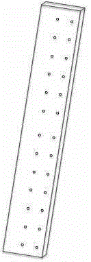
[0030] The costal cartilage tissue of freshly slaughtered pigs was cleaned, the periosteum was removed, cut into strips with a thickness of 0.5-4 cm, and washed 3 times with water for injection.

[0031] (2) Virus inactivation

[0032]Adopt low-concentration peracetic acid-ethanol solution method to inactivate virus, this step is carried out in the constant temperature ultrasonic cleaning device that cleaning tank can vibrate, and wherein the volume percentage composition of peracetic acid is 0.05~0.2% (preferably 0.1%), The inactivation time is 1-2h (preferably 1h), the temperature range is 4-40°C, and then washed 2-5 times in phosphate buffered saline, each time for 15min, and the pH value of the washed phosphate buffered saline is detected, When the pH reaches 6.5-7.5, wash the material with flowing water for injection, and...

Embodiment 2

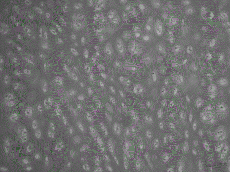
[0041] Embodiment 2: the physical performance, chemical performance, histology and biological performance detection of the medical cartilage scaffold material prepared in embodiment 1

[0042] 1. Physical performance testing

[0043] 1) Mechanical performance testing

[0044] Preparation of the materials provided in Example 1, the materials prepared by Courtman's four-step method and the natural porcine costal cartilage tissue without decellularization, each group of materials took 3 samples, soaked in phosphate buffer saline for 30 minutes, and used The multi-functional mechanical measuring instrument detects the compressive modulus, and calculates the compressive modulus according to the formula. Compression modulus=ΔP / A×L / ΔL (ΔP represents the pressure difference between two points in the linear segment of the pressure-displacement curve, A represents the area of ​​the material under test, L represents the thickness of the material, and ΔL represents the displacement betwe...


PUM

PropertyMeasurementUnit
compressive modulusaaaaaaaaaa

Abstract

The invention provides a preparation method of a medical cartilage support material. The preparation method comprises the following operation steps of: pretreatment, separation and primary washing of a cartilage tissue, inactivation of virus, decellularization, sodium chloride treatment, forming, and packaging and sterilization. The bone marrow-derived cell component and DNA (Deoxyribonucleic Acid) component of an animal are completely removed from the medical cartilage support material prepared by the method, meanwhile a natural ECM (Extracellular Cell Matrix) component and a three-dimensional are completely remained, and no endotoxin, organic solvent and toxic solvent are left; the medical cartilage support material has a porosity suitable for cell ingrowth and cartilage tissue regeneration, and also has ideal mechanical properties.

Description

technical field [0001] The invention relates to the technical field of medical biomaterials, in particular to a method for preparing a medical cartilage scaffold material. Background technique [0002] Cartilage tissue is composed of chondrocytes and cartilage matrix, which is composed of macromolecules such as collagen and cartilage mucin. Cartilage tissue is distributed on the articular surface, ear, nose, intervertebral disc, tracheal ring, etc., and its function is to maintain the normal function of the joint and support the shape and function of the organ. Since there is no blood vessel distribution in the cartilage, and chondrocytes are surrounded by surrounding matrix components, the self-repair ability of cartilage damage is extremely poor, especially for articular cartilage damage, and irreversible pathological processes usually occur, causing changes in joint function. How to effectively repair damaged articular cartilage is a well-known problem. The source of au...

Claims


Application Information

Patent Timeline
Patent Type & Authority Patents(China)
IPC IPC(8): A61L27/36A61L27/56A61L27/54
Inventor 赵博王振军
Owner BEIJING BIOSIS HEALING BIOLOGICAL TECH
Why Eureka
Try Eureka