Recombinant phage double expression vector and application

A bacteriophage and double expression technology, applied in the field of recombinant bacteriophage double expression vectors, can solve the problems of unsatisfactory immune effect and the like, and achieve the effects of convenient large-scale production, low cost and simple preparation method

Active Publication Date: 2015-03-11
JIANGSU ACAD OF AGRI SCI
View PDF5 Cites 0 Cited by
  • Summary
  • Abstract
  • Description
  • Claims
  • Application Information

AI Technical Summary

Problems solved by technology

[0003] However, the immune effect of vaccines prepared with phage particles displaying antigenic epitopes on the surface as antigens is not satisfactory.

Method used


Image

  • Recombinant phage double expression vector and application
  • Recombinant phage double expression vector and application
  • Recombinant phage double expression vector and application

Examples


Embodiment 1

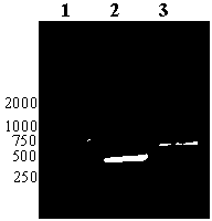
[0035] Example 1 Construction of recombinant phage T7-VP1 129-169

[0036] recombinant phage T7-VP1 129-169 It is formed by inserting the nucleotide sequence encoding the VP1 structural protein (129-169) polypeptide downstream of the p10B gene of T7 Select 415-1b, and the carboxy-terminal of the capsid protein p10B is fused to express the VP1 structural protein of the foot-and-mouth disease virus (129-169) position polypeptide.

[0037] Polypeptide A is formed by adding a flexible Linker amino acid GGGGS to the amino-terminus of the polypeptide at position (129-169) of the VP1 structural protein of foot-and-mouth disease virus (as shown in SEQ ID NO:3). A restriction site EcoRI was added to the 5' end of the nucleotide sequence encoding polypeptide A, and a restriction site Hind III was added to the 3' end to form the DNA sequence shown in SEQ ID NO: 1.

[0038] The DNA sequence shown as SEQ ID NO: 1 was synthesized by a commercial company, and cloned into the multiple clon...

Embodiment 2

[0054] Example 2 Construction of Homologous Recombination Intermediate Plasmid Vector

[0055] Analysis of recombinant phage T7-VP1 129-169 For the genome, the 578th position A on the left side of the genome was selected as the insertion site for the eukaryotic expression cassette. recombinant phage T7-VP1 129-169 The gene fragment at positions 1-578 on the left side of the genome is used as the left homology arm, and the gene segment at positions 2746-2946 on the left side of the genome is the right homology arm. Two pairs of primers were designed for the two homology arms, and XhoI, HindIII, EcoRI, BamHI restriction sites were added at the 5' end of the primers. The left homology arm primers are L1-578UP and L1-578DOWN, and the right homology arm primers are R2746-2946UP and R2746-2946DOWN.

[0056] L1-578UP: 5'-aactcgagTCTCACAGTGTACGGACCTA,

[0057] L1-578DOWN: 5'-AAAAGCTTTCGTGCGACTTATCAGGCTG.

[0058] R2746-2946UP: 5'-AAGAATTCcaGAATTCCAGAAAGAAATTGACCGCGC,

[0059] R...

Embodiment 3

[0076] Example 3 Construction of recombinant T7 phage inserted into eukaryotic expression cassette

[0077] Homologous recombination plasmid pBluescript-L-VP1-R and recombinant phage T7-VP1 129-169 After DNA homologous recombination, the eukaryotic expression cassette composed of CMV eukaryotic promoter, foot-and-mouth disease virus VP1 structural protein gene sequence and SV40 polyA was inserted into the recombinant phage T7-VP1 129-169 After the 578th A on the left side of the genome, the recombinant phage T7-VP1 was replaced 129-169 Fragment at position 579-2745 in the genome to obtain recombinant phage T7-VP1 129-169 -VP1.

[0078] The specific method of homologous recombination is as follows: import the intermediate vector pBluescript-L-VP1-R into E. coli In BL21, BL21-pBluescript-L-VP1-R was obtained. Streak inoculate BL21-pBluescript-L-VP1-R on the LB solid medium plane containing ampicillin resistance, culture overnight at 37°C; pick a single colony from the plat...


PUM


Abstract

The invention provides a recombinant phage double expression vector and application, relating to the technical field of biology. The recombinant phage double expression vector is a recombinant double expression vector obtained after inserting a target gene 1 into the downstream part of a gene p10B in a T7 phage genome and inserting a eukaryotic expression cassette containing a target gene 2 into a non-coding region in the T7 phage genome. The recombinant phage double expression vector has high stability, can display the protein coded by the target gene 1 on the surface of the phage, simultaneously can transfer the eukaryotic expression cassette into an immune cell to achieve eukaryotic expression of the target gene 2, and can serve as a common platform for expression of various epitopes, namely polypeptides. Immune efficacy detection finds that bodies can generate high-level VP1 structural protein antibodies against foot and mouth disease viruses after being immunized by the vaccine, and the antibodies have long duration.

Description

technical field [0001] The invention relates to the field of biotechnology, in particular to a recombinant phage double expression vector and its application. Background technique [0002] T7 phage is a small and virulent phage that infects Escherichia coli, and its genome is a linear double-stranded DNA with a length of 39 936 bp. Its capsid proteins include major noggin (P10A), minor noggin (P10B), collarin (P8), tail protein (P11, P12) and tail protein (P17), among which P10A and P10B are encoded by p10 It is a pair of overlapping proteins, P10A has 344 amino acid residues, and P10B has 397 amino acid residues, which are produced by a translational frameshift at phe341 of P10A. In wild-type T7 phage, P10B accounts for about the total amount of capsid protein 10% of (415 copies). The T7 phage display system is a new type of display system, p10 has been modified, the translation frameshift site in the natural state is removed, and a multiple cloning site is recombined aft...

Claims


Application Information

Patent Timeline
Patent Type & Authority Patents(China)
IPC IPC(8): C12N15/63C12N7/01A61K39/135A61P31/14
Inventor 徐海王义伟鲍熹侯继波
Owner JIANGSU ACAD OF AGRI SCI
Why Eureka
Try Eureka