Method for detecting valence of antibody
A detection method and technology of antibody titer, applied in chemical instruments and methods, biological testing, material testing products, etc., can solve the problems of immature detection methods and long detection time, and achieve good reactivity, good linearity, and reaction conditions. mild effect
- Summary
- Abstract
- Description
- Claims
- Application Information
AI Technical Summary
Problems solved by technology
Method used
Image
Examples
preparation example Construction
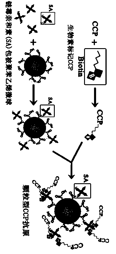
[0040] Preparation of streptavidin-labeled polystyrene microspheres
[0041] 1) Take 0.1mL carboxylated latex microspheres, add 0.9mL ethanesulfonic acid buffer (MES) and mix well, then add water-soluble carbodiimide cross-linking agent (EDC) and N-hydroxysuccinimide (NHS) Activation, EDC concentration 1.0-1.5g / L, ratio of EDC to NHS 1:1-1:2, gentle stirring at room temperature, time controlled at 15-30min;
[0042] 2) Wash by centrifugation, centrifuge at 12000-15000g for 10-30min, wash the pellet three times with 0.1mol / L phosphate buffer (PBS), remove the supernatant, resuspend the pellet in 0.1mol / L PBS, oscillate, and sonicate Promptly obtain the activated latex microsphere;
[0043] 3) Dissolve 2 mg of streptavidin in PBS buffer with pH 6.5-7.8, add it to 1 mL of activated latex microsphere solution, stir gently at room temperature, and react for 2-4 hours;
[0044] 4) After the reaction time is reached, centrifuge at 12000-15000g for 10-30min, precipitate the micros...
Embodiment 1
[0048] 1) Dissolving CCP polypeptide: the prepared CCP polypeptide was dissolved in 0.15M pH9.0 phosphate buffer and stored at 4°C;
[0049] 2) Dissolving esterified biotin: dissolve esterified biotin with dimethyl sulfoxide (DMSO);
[0050] 3) Modification: quickly mix the dissolved CCP polypeptide solution with the dissolved esterified biotin solution, the ratio of biotin to CCP is 10:1, stir and incubate at room temperature for 4 hours;
[0051] 4) Remove excess biotin with a desalting column;
[0052] 5) Elute the sample with 0.05M PBS buffer, pH7.6.
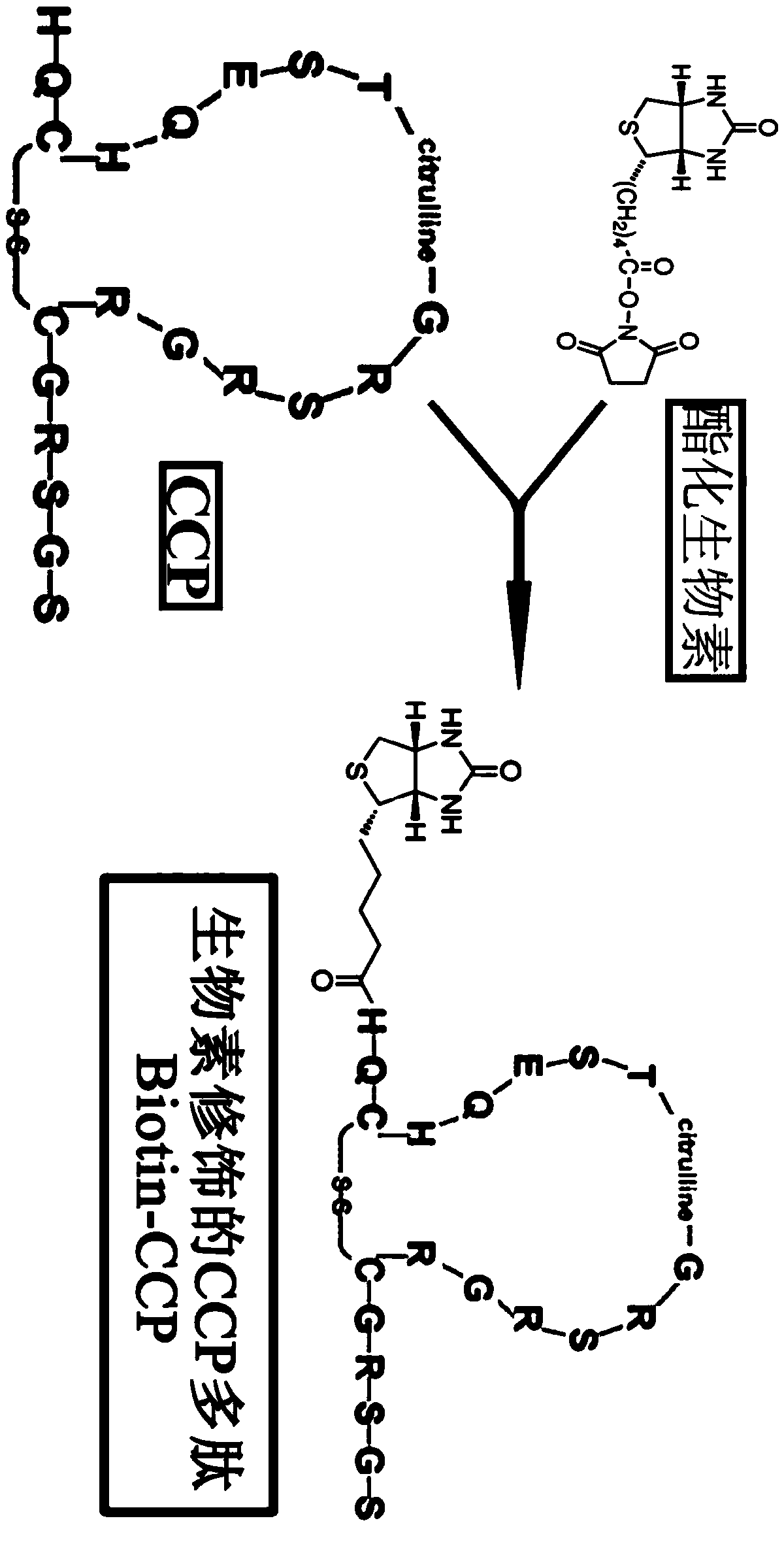
[0053]Collect the eluate and evaluate the labeling result according to the UV absorption spectrum: According to the comparison of the UV absorption spectrum between the biotin-labeled CCP product and the unbiotin-labeled CCP at the same concentration (1mg / mL), the biotin-CCP between 210nm-230nm Increased absorbance values, such as image 3 . ELISA method was used to identify the effect of biotin-labeled CCP: the prepared...
Embodiment 2
[0056] 1) Dissolving CCP polypeptide: the prepared CCP polypeptide was dissolved in 0.2M pH9.6 phosphate buffer and stored at 4°C;
[0057] 2) Dissolving esterified biotin: dissolve esterified biotin with dimethyl sulfoxide (DMSO);
[0058] 3) Modification: quickly mix the dissolved CCP polypeptide solution with the dissolved esterified biotin solution, and the ratio of biotin to CCP is 10:1. Stir and incubate at room temperature for 4 h;
[0059] 4) Remove excess biotin with a desalting column;
[0060] 5) Elute the sample with 0.1M PBS buffer, pH7.2.
[0061] Collect the eluate and evaluate the labeling result according to the UV absorption spectrum: According to the comparison of the UV absorption spectrum between the biotin-labeled CCP product and the unbiotin-labeled CCP at the same concentration (1mg / mL), the biotin-CCP between 200nm-230nm Increased absorbance values, such as Figure 5 .
[0062] ELISA method was used to identify the effect of biotin-labeled CCP: th...
PUM
 Abstract
Description
Claims
Application Information
 - R&D
- Intellectual Property
- Life Sciences
- Materials
- Tech Scout
- Unparalleled Data Quality
- Higher Quality Content
- 60% Fewer Hallucinations
Browse by: Latest US Patents, China's latest patents, Technical Efficacy Thesaurus, Application Domain, Technology Topic, Popular Technical Reports.
© 2025 PatSnap. All rights reserved.Legal|Privacy policy|Modern Slavery Act Transparency Statement|Sitemap|About US| Contact US: help@patsnap.com


