Method for separating exosome from cell culture medium

A technology for separating cells and culturing cells, which is applied in the fields of biochemistry and cell biology, and can solve the problems of high price, impact on experiments, and large consumption of kits, and achieve the effect of saving costs and reducing dosage

Inactive Publication Date: 2013-12-25
SHANXI UNIV
View PDF3 Cites 23 Cited by
  • Summary
  • Abstract
  • Description
  • Claims
  • Application Information

AI Technical Summary

Problems solved by technology

[0004] The first is ultra-high-speed centrifugation or density gradient centrifugation. This method has a limiting factor, that is, it needs to be equipped with an ultra-high-speed centrifuge, and the price of this equipment is very expensive, which limits some small and medium-sized laboratories. secretory research
[0005] The second is to use a kit to extract, this method also has disadvantages, when using this method, the collected fresh medium, only through low centrifugal force to r

Method used


Image

  • Method for separating exosome from cell culture medium
  • Method for separating exosome from cell culture medium
  • Method for separating exosome from cell culture medium

Examples


Embodiment 1

[0022] The colon cancer cells DLD1 in the logarithmic growth phase were inoculated in three cell culture dishes with a diameter of 10 cm, and each dish was 7×10 5 cells at 37°C, 5% CO 2 Cultured in RPMI1640+10%FBS culture medium without antibiotics under the condition of RPMI1640+10%FBS culture medium without antibiotics, when it grew to a confluence of 90%, replaced with new RPMI1640+10%FBS cell culture medium without antibiotics, 10mL per plate, cultured After 24 hours, collect about 30mL of the medium in the three dishes into two 50mL centrifuge tubes, and then number them A and B respectively. Methods to isolate exosomes.

[0023] The inventive method: the steps are as follows:

[0024] 1) Collect fresh cell culture-based centrifuge tubes, centrifuge at 300g for 15 minutes at 4°C to remove suspended cells and cell debris in the medium.

[0025] 2) Transfer the supernatant after centrifugation in step 1) into a new centrifuge tube, and centrifuge at 10,000g for 30 minute...

Embodiment 2

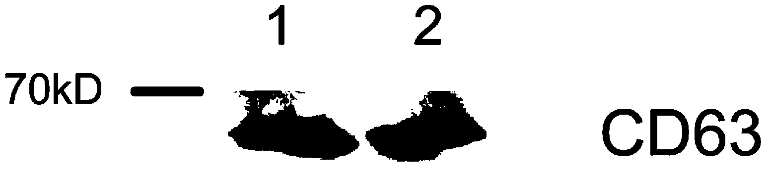
[0030] The exosomes isolated in Example 1 were used to detect the marker protein CD63 in the exosomes by western blot test.

[0031] (1) Add an appropriate amount of membrane protein extract containing PMSF to the exosomes isolated above, incubate at 4°C for 30 min, and then repeatedly freeze and thaw in liquid nitrogen three times to fully lyse them.

[0032] (2) Determine the protein concentration in the lysate by using the BCA method to ensure that the total amount of protein in each sample is the same, then add 5× loading buffer, denature at 100°C for 5 minutes, and perform 10% SDS-polyacrylamide gel electrophoresis. After electrophoresis, the protein was transferred to PVDF membrane by wet transfer method, and blocked with 5% skimmed milk powder for 2h.

[0033] (3) After blocking, wash off the blocking solution with TBST buffer, add CD63 antibody, and incubate overnight at 4°C. The next day, after washing away the primary antibody with TBST, add horseradish peroxidase-l...

Embodiment 3

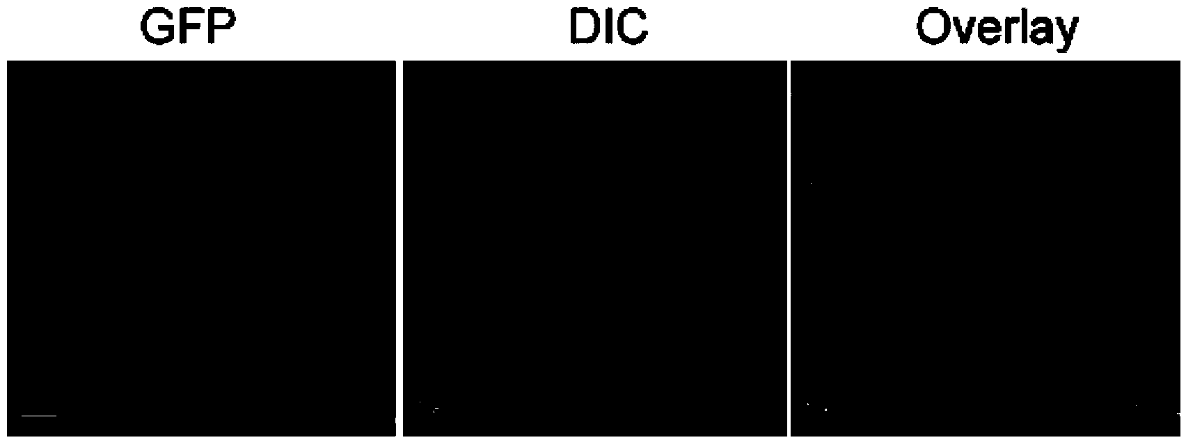
[0036] The exosomes isolated by the method of the present invention were observed by a fluorescence microscope.

[0037] (1) Resuspend the exosomes isolated in Example 1 with an appropriate amount of PBS, add DiO (green fluorescent probe for cell membrane), and stain for about 10 minutes, so that the exosomes are marked with green fluorescence.

[0038] (2) Centrifuge at 10000g for 1 hour, discard the supernatant and add PBS to wash away unbound probe molecules.

[0039] (3) Repeat the step in (2) once.

[0040] (4) Re-suspend the isolated exosomes in PBS, smear the suspension on a glass slide, seal the slide after drying, and observe under a fluorescence microscope.

[0041] The result is as image 3 The isolated exosomes shown were stained green, and the diameters of the exosomes were consistent with those reported in the literature.


PUM


Abstract

The invention provides a method for separating an exosome from a cell culture medium. The method comprises the following steps of: collecting fresh cell culture medium, centrifuging the collected cell culture medium to remove the suspended cell, other cell fragments and impurities in the culture medium, transferring the supernatant into a concentration tube for concentration, and adding Total Exosome Isolation (invitrogen) into the concentrated culture medium for the final extraction of the exosome. By adopting the method provided by the invention, the exosome in the cell culture medium can be conveniently obtained, and the separation cost is greatly reduced.

Description

technical field [0001] The invention relates to the fields of biochemistry and cell biology, in particular to a method for isolating exosomes in a cell culture medium. Background technique [0002] Exosomes are membranous vesicles with a diameter of about 40-100 nm released into the extracellular matrix after the multivesicular body (MVB) in the cell fuses with the cell membrane. Many cells can secrete exosomes, such as T cells, B cells, dendritic cells, mast cells, and tumor cells. Exosomes contain a variety of proteins and RNAs, which transmit information and regulate each other between cells, but the way of transmitting information and the mechanism of mutual regulation are not completely clear, especially between tumor cells : Do exosomes transfer the characteristic information of cancer cells to paracancerous tissues, and promote their carcinogenesis through the contained proteins? Do exosomes affect the migration, proliferation and invasion of tumor cells? and many ...

Claims


Application Information

Patent Timeline
IPC IPC(8): C12N5/09C12N5/0781C12N5/0783C12N5/0784C12N5/078
Inventor 张立超李宗伟李卓玉李汗卿
Owner SHANXI UNIV
Why Eureka
Try Eureka