Molecular signature of pigment spots on the skin, associated with extracellular matrix organization
An external matrix, skin technology, applied in the field of pigmentation and the treatment of these pigmentations, can solve the problems of molecular imbalance or dysfunction, and no discussion of pigmentation mechanism dysfunction and other problems
- Summary
- Abstract
- Description
- Claims
- Application Information
AI Technical Summary
Problems solved by technology
Method used
Image

Examples
Embodiment 1
[0200] Example 1: Transcription Studies
[0201] Comparative study of gene expression levels in solar lentigo (LS) damaged skin and adjacent undamaged skin (US).
[0202] The aim of this study was to identify relevant, reproducible and significant markers that reflect the changes associated with solar lentigo formation in order to use them as targets for effective treatment or as a tool for analyzing the efficacy of a given treatment. landmark.
[0203] Briefly, 15 female volunteers were recruited to participate in a "genome-wide" transcriptome study (Array, Inc., USA). For each volunteer, a solar lentigo type lesion was diagnosed on the back of his hand, and this solar lentigo diagnosis was further confirmed by epidermal luminescence microscopy. This check is for:
[0204] 1) Identify lesions (solar lentigo specific) using criteria at the dermal-epidermal junction ("fingerprints") to differentiate lentigo (lack of fingerprints, uniform pigmentation, and moth-eaten border...
Embodiment 2
[0221] Example 2: Materials and methods
[0222] Fifteen female volunteers with phenotypes II-IV, aged between 50 and 70 years, were selected. Their solar lentigines on the dorsum of the hands were at least 3 mm in size. They were characterized by epiluminescence microscopy. Various advantages of characterization by epiluminescence microscopy have been indicated in Example 1.
[0223] Two 3 mm diameter biopsies were taken from one hand of each patient. For each volunteer, one biopsy corresponded to the solar lentigines lesion (LS) and the other corresponded to the other adjacent undamaged skin (US) (again confirmed by epidermal luminescence microscopy).
[0224] 3mm biopsy sections were sampled and placed in RNAlater (Qiagen cat. 76106) at 4°C for 16 to 24 hours. The next day, samples were placed at -20°C pending the homogenization and extraction steps. After thawing, samples were minced with a scalpel to facilitate homogenization prior to transfer to lysis buffer.
[...
Embodiment 3
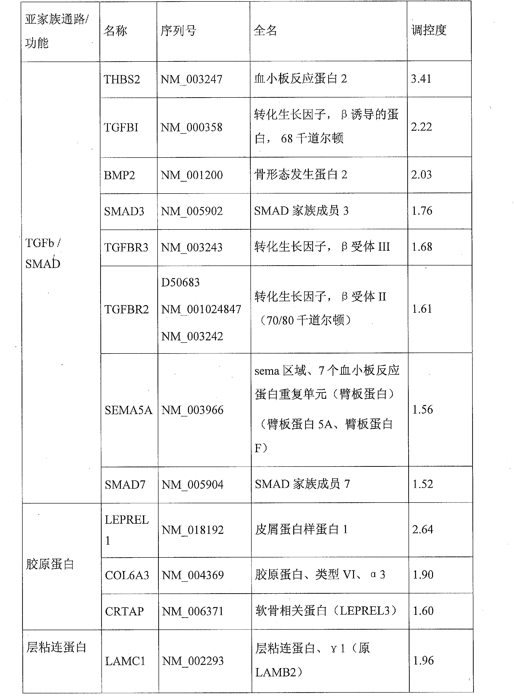
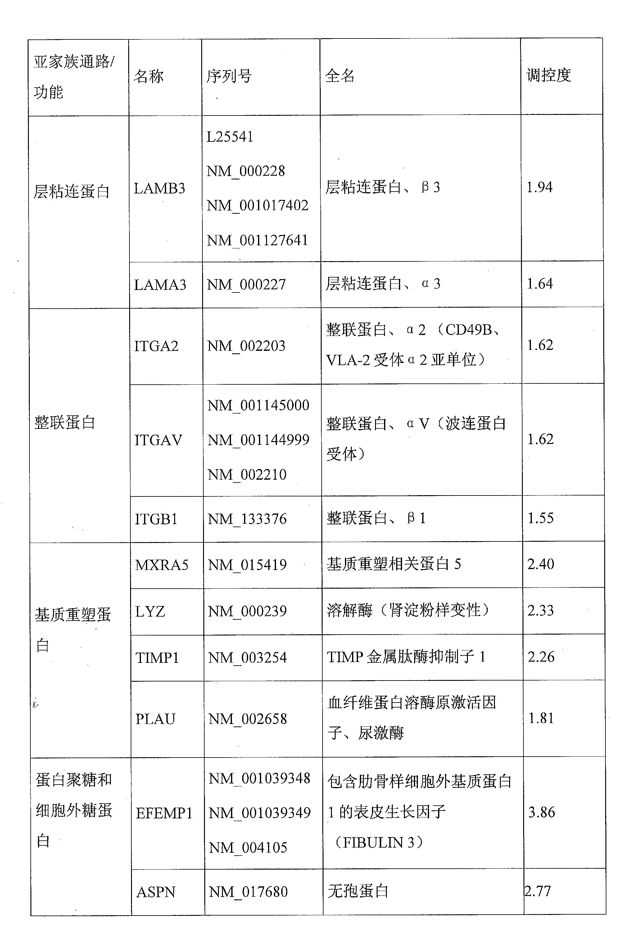
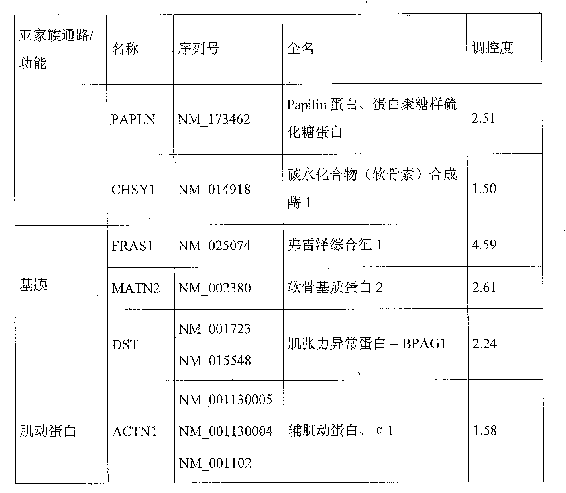
[0243] Embodiment 3: Regulator:
[0244] There are compounds known to modulate protein dysregulation in lentigo. The following can be used as extracellular matrix gene / protein families:
[0245] - Fibrates including fenofibrate, for example to reduce the expression of SMAD3,
[0246] - alkaloids including oxymatrine, which reduces SMAD3, or polyphenols, such as salvianolic acid B, which reduces the expression of SMAD3 and TGFBR2,
[0247] - Natural extracts, especially herbal medicines such as Wenpi Decoction and Wuling Powder extract, which can reduce the expression of SMAD3,
[0248] - compounds of the imidazole family, which are antagonists of TGF-βR1, such as SB-431542, which reduces the expression of SMAD3,
[0249] - certain miRNAs, in particular miR183 and miR29b, which reduce the expression of ITGB1,
[0250] - inhibitors of urokinase-type plasminogen activators (uPA), such as p-aminobenzamidine or B428 4-substituted benzo[b]thiophene-2-carboxamidines, which redu...
PUM
 Abstract
Description
Claims
Application Information
 - R&D
- Intellectual Property
- Life Sciences
- Materials
- Tech Scout
- Unparalleled Data Quality
- Higher Quality Content
- 60% Fewer Hallucinations
Browse by: Latest US Patents, China's latest patents, Technical Efficacy Thesaurus, Application Domain, Technology Topic, Popular Technical Reports.
© 2025 PatSnap. All rights reserved.Legal|Privacy policy|Modern Slavery Act Transparency Statement|Sitemap|About US| Contact US: help@patsnap.com