Internal rectal optical, optoacoustic and ultrasonic multimode imaging endoscope and imaging method thereof

A multi-mode imaging and endoscopy technology, applied in endoscopy, medical science, diagnosis, etc., can solve the problems of insufficient imaging resolution and inability to provide detailed information of rectal wall and sub-rectal tissue

Active Publication Date: 2014-04-02
广州佰奥廷电子科技有限公司
View PDF8 Cites 39 Cited by
  • Summary
  • Abstract
  • Description
  • Claims
  • Application Information

AI Technical Summary

Problems solved by technology

[0003] Existing methods include rectal electronic endoscopy and rectal ultrasonography. Although rectal electronic endoscopy can clearly show the external shape of the rectal inner wall, it cannot provide detailed information on the rectal wall and rectal submural tissue; rectal ultrasonography Endoscopic imaging is based on detecting the echoes of ultrasound signals in the rectal wall. It can image the tissues under the rectal wall and reflect the differences in the acoustic impedance of rectal tissues, but the imaging resolution is not enough
[0004] The prior art "A Photoacoustic Endoscopic Imaging Method and Its Device" discloses a photoacoustic endoscopic imaging detection method and its device. The photoacoustic endoscopic imaging technology is based on the photoacoustic signal generated after the tissue absorbs the pulse laser Imaging is performed to reflect the difference in optical absorption within the tissue, combining the advantages of high contrast characteristics of pure optical imaging and high penetration depth characteristics of pure ultrasound imaging, but photoacoustic imaging can only reflect the single parameter information of tissue optical absorption difference

Method used


Image

  • Internal rectal optical, optoacoustic and ultrasonic multimode imaging endoscope and imaging method thereof
  • Internal rectal optical, optoacoustic and ultrasonic multimode imaging endoscope and imaging method thereof
  • Internal rectal optical, optoacoustic and ultrasonic multimode imaging endoscope and imaging method thereof

Examples


Embodiment Construction

[0053] The present invention will be further described below in conjunction with drawings and embodiments.

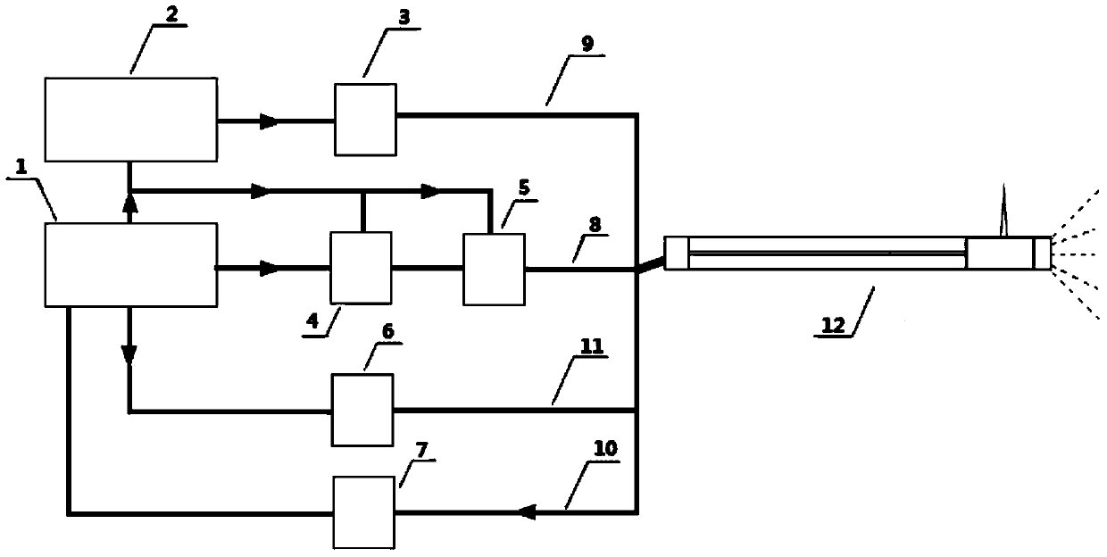
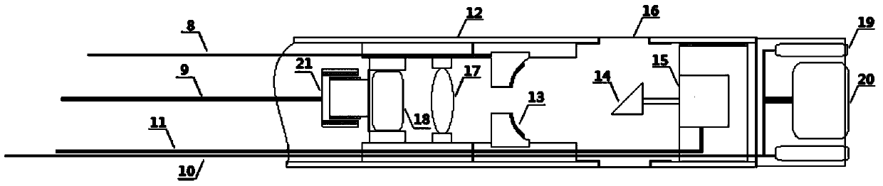
[0054] Such as figure 1 The shown rectal optical, photoacoustic, ultrasonic multimode imaging endoscope and its imaging method include a cannula 12, a photoacoustic signal excitation component, an ultrasonic signal excitation and acquisition component, an optical imaging component, and an image reconstruction and display component;

[0055] The photoacoustic signal excitation component includes a pulsed laser 2, a single-mode fiber 9, a fiber-coupled collimator 3 and an achromatic focusing lens 17;

[0056] The ultrasonic signal excitation and collection components include an ultrasonic pulse transmitter receiver 5, a fiber optic collimator 18, a hollow focused ultrasonic transducer 13, a coated acoustic / optical high reflector 14, a micro stepper motor 15 and an imaging window 16;

[0057] The optical imaging assembly includes a miniature adjustable focus optical camer...


PUM


Abstract

The invention discloses an internal rectal optical, optoacoustic and ultrasonic multimode imaging endoscope which comprises a sleeve, an optoacoustic signal activating component, an ultrasonic signal activating and collecting component, an optical imaging component and an image reconstructing and displaying component. A device comprises a compact internal rectal optical, optoacoustic and ultrasonic multimode imaging endoscope, three kinds of imaging can be performed simultaneously, multi-parameter physical information and multi-scale structural images in recta can be acquired. The invention further provides a configuration scheme of the whole device and a method for utilizing the device for imaging. Three imaging techniques are highly integrated into one set of instrument, each imaging technique is optimized, and combination of three rectal endoscopic imaging methods of optical imaging, optoacoustic imaging and ultrasonic imaging is realized. The integrated endoscope combines specific advantages of three imaging modes, rectal physical images with multi-parameter information and multi-scale structural characteristics can be acquired, and application requirements of a rectal endoscope on medicine can be met better.

Description

technical field [0001] The invention belongs to the technical field of endoscope non-destructive testing, and in particular relates to an endoscope device and an imaging method integrating optical, photoacoustic and ultrasonic multi-mode imaging in the rectum. Background technique [0002] With the popularization and application of endoscopes in medicine, it is particularly important to design and manufacture more modern, rationalized, humanized and intelligent endoscopes. The application of rectal endoscopy plays a very important role. [0003] Existing methods include rectal electronic endoscopy and rectal ultrasonography. Although rectal electronic endoscopy can clearly show the external shape of the rectal inner wall, it cannot provide detailed information on the rectal wall and rectal submural tissue; rectal ultrasonography Endoscopic imaging is based on detecting the echoes of ultrasound signals in the rectal wall. It can image the tissues under the rectal wall and re...

Claims


Application Information

Patent Timeline
Patent Type & Authority Applications(China)
IPC IPC(8): A61B1/04
Inventor 杨思华刘宁邢达
Owner 广州佰奥廷电子科技有限公司
Why Eureka
Try Eureka