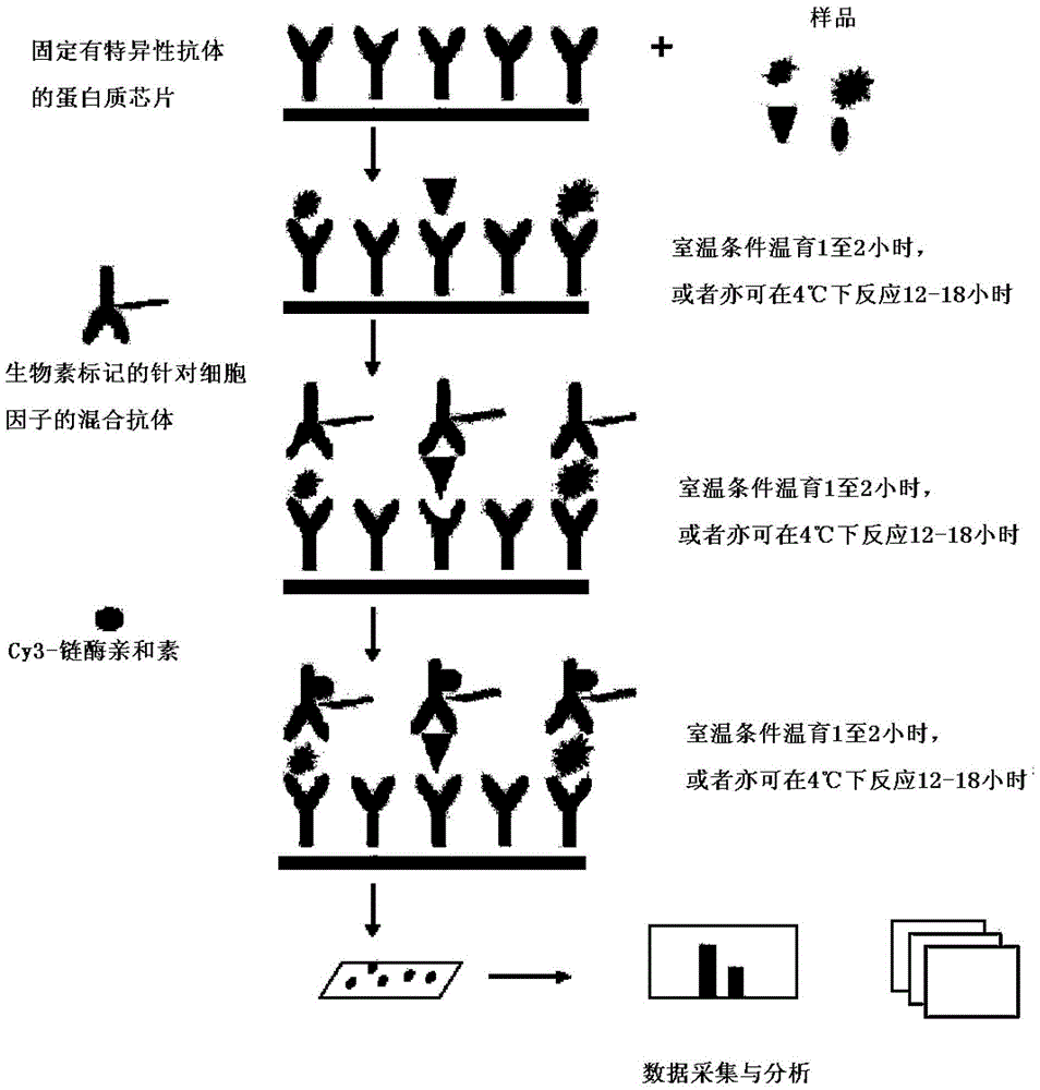
An improved antibody chip kit for simultaneous quantitative detection of multiple cytokines
A cytokine and quantitative detection technology, applied in the field of biomedicine, can solve the problems of cumbersome operation, low sample consumption, low sensitivity, etc., and achieve the effects of increasing stability, reducing background interference, and increasing detection sensitivity.
- Summary
- Abstract
- Description
- Claims
- Application Information
AI Technical Summary
Problems solved by technology
Method used
Image
Examples
Embodiment 1
[0031] Example 1: Screening of the best antibody chip carrier.
[0032] Conventional antibody chips mostly use nitrocellulose membranes as carriers. Because the nitrocellulose membranes are multi-layered structures, it is difficult to wash the chips, so the results of the chips fluctuate greatly. At the same time, because the nitrocellulose membrane chip is not easy to operate on a large scale, the use of large-scale clinical samples is not yet common. Different manufacturers use different methods to fix the nitrocellulose membrane on the surface of the glass slide. Among them, Whatman's SS glass slides, which fix 16 cells containing nitrocellulose membrane on one glass slide, and Gentel has processed it. PATH slides were produced by coating the surface of the slide with nitrocellulose material. In addition, in order to coat the antibody on the slide surface, we screened the carriers activated by different methods. Cy3 and Cy5-labeled streptavidin was spotted on the surface ...
Embodiment 2
[0034] Example 2: Preparation of an antibody chip kit for simultaneous quantitative detection of multiple cytokines.
[0035] In order to detect the presence of the corresponding cytokines in the samples, prepare slides immobilized with specific antibodies against the following proteins: granulocyte-macrophage colony stimulating factor (GM-CSF), interferon gamma (IFNgamma), interleukin 2 (IL-2), Interleukin 4 (IL-4), Interleukin 5 (IL-5), Interleukin 6 (IL-6), Interleukin 8 (IL-8), Interleukin 10 (IL-10), Interleukin 13 (IL -13), tumor necrosis factor alpha (TNFalpha).
[0036] 1. Antibody preparation:
[0037] Specific antibodies against the proteins listed in Table 1 are used. The source, concentration, and protein names of the antibodies are detailed in Table 1:
[0038] Table 1 Name of the antigenic protein targeted by the specific antibody, source and concentration information of the antibody
[0039]
[0040]
[0041] 2. Preparation and storage of antibody chips...
Embodiment 3
[0051] Example 3: Experiment for quantitative detection of cytokines using the kit of the present invention.
[0052] 1. Complete drying of slide chips
[0053] Take the slide chip out of the box, and after equilibrating at room temperature for 20-30min, open the packaging bag, peel off the seal, and then place the chip in a vacuum desiccator or dry at room temperature for 1-2 hours.
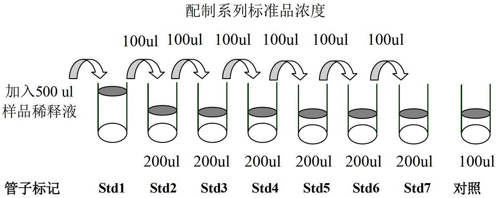
[0054] 2. Press image 3 Cytokine standard gradients were serially diluted as shown in
[0055] 2.1. Add 500 μl of sample diluent to the vial of cytokine standard mixture to re-dissolve the standard. Before opening the vial, centrifuge quickly, gently beat up and down to dissolve the powder, and mark this vial as Std1.
[0056] 2.2. Label 6 clean centrifuge tubes as Std2, Std3 to Std7, and add 200 μl of sample diluent to each tube.
[0057] 2.3. Add 100 μl of Std1 to Std2 and mix gently, then extract 100 μl from Std2 and add it to Std3, and dilute to Std7 in this way.
[0058] 2.4. Take 100...
PUM
 Abstract
Description
Claims
Application Information
 - R&D
- Intellectual Property
- Life Sciences
- Materials
- Tech Scout
- Unparalleled Data Quality
- Higher Quality Content
- 60% Fewer Hallucinations
Browse by: Latest US Patents, China's latest patents, Technical Efficacy Thesaurus, Application Domain, Technology Topic, Popular Technical Reports.
© 2025 PatSnap. All rights reserved.Legal|Privacy policy|Modern Slavery Act Transparency Statement|Sitemap|About US| Contact US: help@patsnap.com


