Preparation method and application of fluorescence-MRI (Magnetic Resonance Imaging) dual-mode image probe

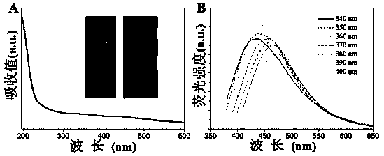
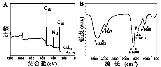
A dual-mode, probe technology, applied in the fields of nanomaterials and medical engineering, can solve the problems of short residence time, liver and kidney toxicity and side effects in clinical applications, and achieve simple reaction equipment, shortened longitudinal relaxation time, and excellent optical properties. Effect

Inactive Publication Date: 2014-11-26
JIANGSU UNIV
View PDF2 Cites 11 Cited by
  • Summary
  • Abstract
  • Description
  • Claims
  • Application Information

AI Technical Summary

Problems solved by technology

However, due to the short residence time in the body, liver and kidney toxicity and other side effects seriously affect its clinical application.

Method used


Image

  • Preparation method and application of fluorescence-MRI (Magnetic Resonance Imaging) dual-mode image probe
  • Preparation method and application of fluorescence-MRI (Magnetic Resonance Imaging) dual-mode image probe
  • Preparation method and application of fluorescence-MRI (Magnetic Resonance Imaging) dual-mode image probe

Examples


Embodiment 1

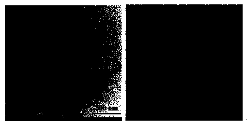
[0029] Dissolve 1 g of glycine and 1 g of gadopentetate meglumine in 20 mL of double-distilled water, and stir magnetically at room temperature to fully dissolve to obtain a uniform and transparent solution. Add the above solution to a household microwave oven (power; 700W) and heat it at high temperature for 1 minute. After the solution cools down naturally, filter it with a medium-speed filter paper to remove insoluble black precipitates. Centrifuge at 12,000 g for 30 minutes to remove large particles. Collect the supernatant and inject it into the Molecular Retention Dialysis was carried out in a dialysis bag with a capacity of 2000 Da. The dialysis time was 72 h, and the water was changed every 12 h. The dialyzed product was rotovaped to obtain a concentrated solution. The concentrated solution is freeze-dried at -50° C. to powder to obtain gadolinium-doped carbon quantum dots.

Embodiment 2

[0031] Dissolve 1 g of glycine and 2 g of gadopentetate meglumine in 20 mL of double-distilled water, and stir magnetically at room temperature to fully dissolve to obtain a uniform and transparent solution. Add the above solution to a household microwave oven (power; 700W) and heat it at high temperature for 4 minutes. After the solution is naturally cooled, filter it with a medium-speed filter paper to remove insoluble black precipitates. Centrifuge at 12,000 g for 30 minutes to remove large particles. Collect the supernatant and inject it into the Molecular Retention Dialysis was carried out in a dialysis bag with a capacity of 2000 Da. The dialysis time was 72 h, and the water was changed every 12 h. The dialyzed product was rotovaped to obtain a concentrated solution. The concentrated solution was freeze-dried to powder at -50°C to obtain gadolinium-doped carbon quantum dots (yield: 0.82%).

Embodiment 3

[0033] Dissolve 1 g of glycine and 3 g of gadopentetate meglumine in 20 mL of double-distilled water, and stir magnetically at room temperature to fully dissolve to obtain a uniform and transparent solution. Add the above solution into a household microwave oven (power; 700W) and heat it at high temperature for 6 minutes. After the solution cools down naturally, filter it with a medium-speed filter paper to remove insoluble black precipitates, centrifuge at 12000 g for 30 minutes to remove large particles, collect the supernatant and inject it into the molecular interception Dialysis was carried out in a dialysis bag with a capacity of 2000 Da. The dialysis time was 72 h, and the water was changed every 12 h. The dialyzed product was rotovaped to obtain a concentrated solution. The concentrated solution was freeze-dried to powder at -50°C to obtain gadolinium-doped carbon quantum dots (yield: 1.08%).


PUM


Abstract

The invention provides a preparation method and application of a fluorescence-MRI (Magnetic Resonance Imaging) dual-mode image probe, and particularly relates to a dual-mode image probe which combines a fluorescent nanoparticle and a paramagnetic contrast agent and a preparation method thereof, belonging to the field of functional materials. The main process of the method comprises the following step of: removing residues and moisture through dialysis and freeze-drying by taking an amino compound as a surface passivation agent and a gadolinium-based complex as a carbon source material and adopting a one-step forming method to obtain a fluorescence-MRI dual-mode gadolinium-doped carbon quantum dot. The method provided by the invention ahs the characteristics of simple process, low cost and easiness for large-scale production. The fluorescence-MRI dual-mode gadolinium-doped carbon quantum dot prepared through the method has the excellent characteristics of higher fluorescent property, higher relaxation efficiency, good biocompatibility and the like, is successfully applied to in-vitro live cell fluorescence imaging and in-vivo magnetic resonance imaging; the fluorescence-MRI dual-mode gadolinium-doped carbon quantum dot synthesized through the method is about to have wide application prospect in the field of biomedicine.

Description

technical field [0001] The invention relates to a preparation method and application of a fluorescence-MRI dual-mode imaging probe, in particular to a fluorescence-MRI dual-mode gadolinium-doped carbon quantum dot prepared by a one-step method and its application in biomedical imaging, belonging to nano materials and medical engineering. Background technique [0002] Magnetic resonance imaging (MRI) is a non-invasive imaging method without damage and ionizing radiation. It is currently the most mature and effective method for disease diagnosis and treatment and surgical guidance. However, due to the low sensitivity, MRI intensifiers (contrast agents) are often needed in clinical MRI diagnosis to improve the imaging effect of lesion tissue physiology and anatomy. These contrast agents usually use paramagnetic substances to affect the relaxation time of water molecules in local tissues in the body, change the signal intensity, and form a contrast with the surrounding tissues ...

Claims


Application Information

Patent Timeline
Patent Type & Authority Applications(China)
IPC IPC(8): A61K49/06A61K49/00G01N21/64G01N15/10
Inventor 杜凤移张丽张礼荣张苗苗华晔龚爱华邵启祥
Owner JIANGSU UNIV
Why Eureka
Try Eureka