Preparation method of green fluorescent carbon spot and application of green fluorescent carbon spot in cell imaging

A technology of green fluorescence and carbon dots, applied in the field of preparation of fluorescent nanomaterials, can solve the problems of complex preparation methods, harsh experimental conditions, environmental hazards, etc., and achieve the effects of simple preparation methods, low cost, and interference avoidance.

Inactive Publication Date: 2015-03-11
SHANXI UNIV
View PDF6 Cites 24 Cited by
  • Summary
  • Abstract
  • Description
  • Claims
  • Application Information

AI Technical Summary

Problems solved by technology

The top-down synthesis method has harsh experimental conditions and complicated preparation methods; most bottom-up synthesis methods use chemical reagents, which are harmful to the environment

Method used


Image

  • Preparation method of green fluorescent carbon spot and application of green fluorescent carbon spot in cell imaging
  • Preparation method of green fluorescent carbon spot and application of green fluorescent carbon spot in cell imaging
  • Preparation method of green fluorescent carbon spot and application of green fluorescent carbon spot in cell imaging

Examples


Embodiment 1

[0039] Preparation of green fluorescent carbon dots using lily petals as carbon source:

[0040] (1) Add 0.5g of lily petal powder to 20mL of deionized water to prepare a mixed solution;

[0041] (2) Transfer the mixed solution obtained in (1) to a hydrothermal reaction kettle, and conduct a hydrothermal reaction at 200° C. for 3 hours;

[0042] (3) The product obtained in (2) was centrifuged at 4000r / min for 10min in a centrifuge, and then dialyzed for 48h with a dialysis bag with a molecular weight cut-off of 500-1000Da to finally obtain a green fluorescent carbon dot solution.

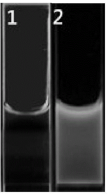
[0043] The photos of the prepared green fluorescent carbon dot solution under the irradiation of fluorescent lamp and ultraviolet lamp with a wavelength of 365nm are shown in figure 1 , wherein 1 is a picture of the green fluorescent carbon dot solution under the irradiation of a fluorescent lamp, and the color is brown, and 2 is a picture of a wavelength of 365nm ultraviolet light, and the color i...

Embodiment 2

[0047] Preparation of green fluorescent carbon dots using rose petals as carbon source:

[0048] (1) 0.5g rose petal powder was added into 20mL deionized water to obtain a mixed solution;

[0049] (2) The mixed solution obtained in (1) was transferred to a hydrothermal reaction kettle, and hydrothermally reacted at 250° C. for 2 h;

[0050] (3) Centrifuge the product obtained in (2) at 4000 r / min for 10 minutes, and then dialyze the product with a dialysis bag with a molecular weight cut-off of 500-1000 Da for 48 hours to finally obtain a green fluorescent carbon dot solution.

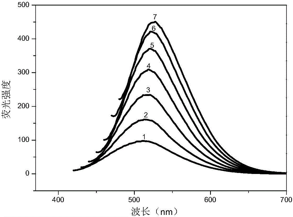
[0051] The fluorescence emission spectra of the prepared green fluorescent carbon dots at different excitation wavelengths are shown in Figure 4 , where 1 to 7 are the fluorescence spectra at excitation wavelengths of 400 nm, 410 nm, 420 nm, 430 nm, 440 nm, 450 nm and 460 nm, respectively.

Embodiment 3

[0053] Preparation of green fluorescent carbon dots using Sophora japonica petals as carbon source:

[0054] (1) 0.5g of Sophora japonica petal powder was added to 30mL of deionized water to prepare a mixed solution;

[0055] (2) The mixed solution obtained in (1) was transferred to a hydrothermal reaction kettle, and hydrothermally reacted at 200° C. for 3 h;

[0056] (3) Centrifuge the product obtained in (2) at 4000 r / min for 10 minutes, and then dialyze the product with a dialysis bag with a molecular weight cut-off of 500-1000 Da for 48 hours to finally obtain a green fluorescent carbon dot solution.

[0057] The fluorescence emission spectra of the prepared green fluorescent carbon dots at different excitation wavelengths are shown in Figure 5 , where 1 to 7 are the fluorescence spectra at excitation wavelengths of 400 nm, 410 nm, 420 nm, 430 nm, 440 nm, 450 nm and 460 nm, respectively.


PUM

PropertyMeasurementUnit
sizeaaaaaaaaaa

Abstract

The invention provides a preparation method of a green fluorescent carbon spot and an application of the green fluorescent carbon spot in cell imaging, and belongs to the preparation and application field of fluorescent nano materials. The preparation method comprises the following steps: (1) adding petal powder into deionized water to prepare a mixed solution; (2) transferring the mixed solution obtained in the step (1) to a hydrothermal reactor to carry out hydrothermal reactions; (3) subjecting the reaction products obtained in the step (2) to centrifugation and dialysis so as to obtain a green fluorescent carbon spot solution. The plant petal is taken as the carbon source, the raw material is easily available, the preparation method is simple, and the cost is low. The prepared green fluorescent carbon spot has the advantages of good water solubility and high stability, and is capable of being directly applied to cell imaging.

Description

technical field [0001] The invention relates to the preparation of fluorescent nanometer materials, in particular to a preparation method of green fluorescent carbon dots and its application in cell imaging. Background technique [0002] Carbon nanodots, also known as carbon dots, are a new class of carbon nanomaterials with a size smaller than 10nm, which were first discovered in 2004 when single-layer carbon nanotubes were purified by electrophoresis. Due to its strong stability, low toxicity, good water solubility, low cost, and abundant raw materials, carbon dots have gradually become a new star in the family of carbon nanomaterials, and are widely used in research fields such as bioimaging, fluorescent printing, and sensing. . [0003] Currently, most of the synthesized fluorescent carbon dots emit blue light when excited by ultraviolet light. In terms of biological imaging, green fluorescent carbon dots are more attractive. This is because the autofluorescence of org...

Claims


Application Information

Patent Timeline
Patent Type & Authority Applications(China)
IPC IPC(8): C09K11/65C01B31/02G01N21/64
Inventor 石利红李艳艳李晓峰温香平双少敏
Owner SHANXI UNIV
Why Eureka
Try Eureka