Primary separation and culture method of human amniotic mesenchymal stem cells

A technique for stromal stem cells and culturing methods, applied in the field of primary separation and culturing of human amniotic mesenchymal stem cells, can solve the problems of decreased activity, long time consumption, low cell yield, etc., and achieves less cell damage, high cell viability, The effect of a large amount of stem cells

Active Publication Date: 2015-03-25
GUANGZHOU SALIAI STEMCELL SCI & TECH CO LTD
View PDF2 Cites 17 Cited by
  • Summary
  • Abstract
  • Description
  • Claims
  • Application Information

AI Technical Summary

Problems solved by technology

The prior art using type II collagenase to separate has disadvanta

Method used


Image

  • Primary separation and culture method of human amniotic mesenchymal stem cells
  • Primary separation and culture method of human amniotic mesenchymal stem cells
  • Primary separation and culture method of human amniotic mesenchymal stem cells

Examples


Embodiment 1

[0051] A primary isolation and culture method of human amniotic mesenchymal stem cells, specifically comprising the following steps:

[0052] 1. Primary isolation of human amniotic mesenchymal stem cells

[0053] Under sterile conditions, the fresh placenta discarded after delivery was placed in a sterile tray, the amniotic membrane was stripped from the placental tissue with surgical instruments, and washed several times with PBS buffer solution to remove residual blood. Cut the amnion tissue into 6×6cm2 in a 15cm glass dish, divide into 50mL centrifuge tubes (each tube contains amnion tissue 10-15cm), add 0.1-0.3% trypsin to 45mL. Transfer the centrifuge tube to a constant temperature shaker at 37°C, 150-300r / min, digest for 30min, and shake vigorously several times every 10min to remove epithelial cells. Transfer the trypsinized amnion tissue to a 250mL storage bottle, add 150mL PBS buffer solution, shake vigorously, and repeat this operation twice. The amnion tissue was ...

example 2

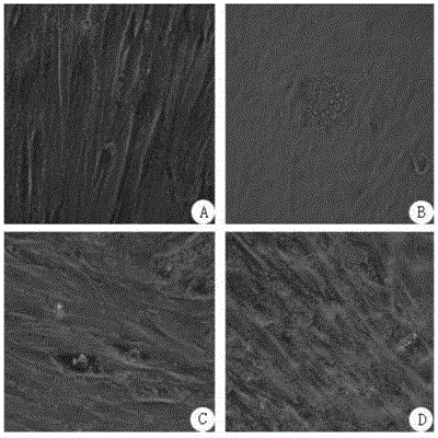
[0057] Flow cytometric identification of isolated amniotic stem cells

[0058] When the primary human amniotic mesenchymal stem cells grow to 80%-90%, collect the cells after trypsinization, take two 1.5ml EP tubes, each tube 1x10 6 Wash two cells with staining buffer (10% FBS+90% PBS), add 200 μl of staining buffer to each tube, add 5 μl of the following four antibodies CD45, CD59, HLADR, and CD90 to the sample, and add no antibody to the negative control. Incubate at 4°C for 20 minutes, wash twice with staining buffer, resuspend with 500 μl 1640 medium, collect 30,000 samples with flow cytometry, and detect the expression of surface markers CD45, CD59, CD90, HLA-DR, etc. . see attached results figure 1 .

[0059] From attached figure 1 It can be seen from the results that the sample expresses CD59 and CD90; low expression of HLA-DR and CD45, which is in line with the characteristics of mesenchymal stem cells. It can also be seen that this method can obtain mesenchymal st...

example 3

[0061] Proliferation curve of isolated stem cells

[0062] The hAMSCs of passage P2 were collected by trypsinization, and the cells were seeded in 96-well plates at 2000 cells / well. at 37°C, 5% CO 2 Continuous culture in the incubator for 7 days. The cells were tested on 1d, 3d, 5d, and 7d respectively. Add 10ul staining agent to the well to be tested, 37°C, 5% CO 2 Cultivate for 2h. The absorbance at 450 nm was measured with a microplate reader, and 6 replicates were made for each sample. The results are shown in Table 1.

[0063] Table 1

[0064]

1

2

3

4

5

6

average

Variance

1d

0.23

0.22

0.2

0.24

0.22

0.21

0.22

0.014142

3d

0.36

0.38

0.38

0.37

0.39

0.36

0.373333

0.012111

5d

0.72

0.69

0.71

0.68

0.69

0.7

0.698333

0.01472

7d

0.82

0.84

0.91

0.88

0.87

0.9

0.87

0.034641

[...


PUM


Abstract

The invention relates to a primary separation and culture method of human amniotic mesenchymal stem cells. The primary separation and culture method of the human amniotic mesenchymal stem cells comprises the following steps of: providing a human amniotic tissue, washing with PBS (phosphate buffer saline), shearing into pieces, adding 0.1-0.3% (by mass) trypsinase to digest, adding I-type collagenase and basal high-glucose DMEM until the final concentration of the I-type collagenase is 0.1-0.2%, digesting, separating the digested human amniotic tissue centrifugally to obtain a precipitate, resuspending the precipitate with PBS, filtering, separating centrifugally, and removing the supernatant to obtain the human amniotic mesenchymal stem cells. Compared with the prior art, the primary separation and culture method provided by the invention has the advantages that because a small amount of trypsinase is used to pretreat and loosen the human amniotic tissue, and further the I-type collagenase with good tissue specificity is used to treat the human amniotic tissue, the digesting time can be shortened greatly, the primary separation steps can be simplified, the human amniotic mesenchymal stem cells can be obtained at a high yield, and the viability of the obtained human amniotic mesenchymal stem cells can be improved greatly compared with that of the human amniotic mesenchymal stem cells in the prior art.

Description

technical field [0001] The invention relates to the technical field of stem cells, in particular to a method for primary isolation and culture of human amniotic mesenchymal stem cells. Background technique [0002] Mesenchymal stem cells (Mesenchymal Stem Cells, MSCs) are mesoderm-derived pluripotent stem cells with high self-renewal ability and multi-lineage differentiation potential. Differentiate into nerve cells, bone cells, chondrocytes, muscle cells, fat cells, etc. under specific conditions. MSCs are pluripotent stem cells with the ability of "horizontal differentiation" or "cross-lineage differentiation". They not only support the growth of hematopoietic stem cells, but also can differentiate into various tissue cells in vitro under different induction conditions. MSCs have broad clinical application prospects and are the preferred seed cells for cell replacement therapy and tissue engineering. [0003] Bone Marrow Derived Mesenchymal Stem Cells (BMSCs) is a kind o...

Claims


Application Information

Patent Timeline
IPC IPC(8): C12N5/0775
Inventor 陈海佳王一飞葛啸虎冯德龙王小燕马岩岩
Owner GUANGZHOU SALIAI STEMCELL SCI & TECH CO LTD
Why Eureka
Try Eureka