

Fluorescent probe method for monitoring Zn<2+> in cancer cells
A fluorescent probe, cancer cell technology, applied in the field of analytical chemistry, can solve many problems and achieve the effects of high luminescence yield, good membrane penetration, and good water solubility
- Summary
- Abstract
- Description
- Claims
- Application Information
AI Technical Summary
Problems solved by technology
Method used
Image

Examples
Embodiment 1
[0037] Embodiment one: the preparation method of each solution, reagent among the present invention
[0038] (1) Preparation of probe solution: Weigh 40.4 mg of probe, dissolve it in DMF, and prepare a 100 mL solution with a concentration of 1.00×10 -3 mol L -1 ; According to the need, gradually dilute with saline to the appropriate concentration.
[0039] (2) Zn 2+ Standard solution: Weigh 59.5mg of analytically pure Zn(NO 3 ) 2 , dissolved in normal saline, and prepared into 100mL solution with a concentration of 2.00×10 -3 mol L -1 ; According to the need, gradually dilute with saline to the appropriate concentration.
[0040] (3) Preparation of other coexisting ionic solutions: Take analytically pure nitrates or hydrochlorides of various metals, dissolve them in double distilled water, and prepare them at a concentration of 2.00×10 -3 mol L -1 double distilled aqueous solution.
[0041] (4) 75% ethanol solution: Add 75mL of absolute ethanol to 100mL with distill...
Embodiment 2
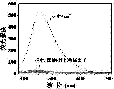
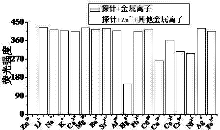
[0048] Embodiment 2: Probe pair Zn 2+Selective fluorescence detection of ions
[0049] Add the DMF stock solution of the probe (1.00×10 -4 mol L -1 , 1mL), metal ion Zn 2+ (2.00×10 -3 mol L -1 , 1 mL), with DMF / H 2 Dilute the O (v / v, 1 / 4) solution to the mark, shake well, and transfer it into a 1cm quartz cuvette for fluorescence spectrum measurement.
[0050] Set the fluorescence excitation wavelength to 360nm, add about 3ml probe (1.00×10 -5 mol L -1 ) DMF / H 2 O (v / v, 1 / 4) solution was tested for fluorescence spectrum, and the probe had no fluorescence emission at a wavelength of 457nm. Join Zn 2+ (2.00×10 -4 mol L -1 ), the probe solution emits strong blue fluorescence. Under the same conditions, Li was added to the probe solution respectively + , Na + , K + , Mg 2+ , Ca 2+ , Ba 2+ ,Sr 2+ , Hg 2+ ,Co 2+ , Ni 2+ , Cu 2+ , Ag + , Pb 2+ , Cd 2+ , Al 3+ , Cr 3+ , Fe 3+ After adding metal ions, the fluorescence spectrum and intensity of the pro...
Embodiment 3
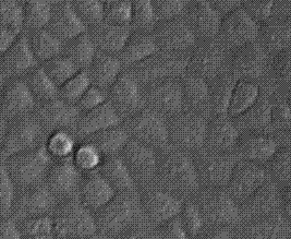
[0052] Example 3: Fluorescence microscopy imaging of living PC3 cells
[0053] (1) Recovery cells
[0054] Take out the PC3 cells from the -80°C refrigerator, place them in 37°C water and shake the cell cryopreservation tube quickly, and thaw them completely within 1-2 minutes. In the aseptic operating table, suck them into the centrifuge tube, and add about 11ml of culture medium (Containing 10% fetal bovine serum, 1% double antibody solution) mix evenly, centrifuge the cell suspension at 1000r / min for 5min, remove the supernatant , Gently blow and break up the cells and culture medium precipitated at the bottom and transfer them to the culture bottle so that the volume of the culture medium in the culture bottle is within 5-7mL, and place it at 37 ℃, containing 5% CO 2 cultured in an incubator.
[0055] (2) Observation → passage → transfer to plate
[0056] Change the culture medium once a day, and observe the cell growth under a microscope until the PC3 cells adhere to t...
PUM
 Abstract
Description
Claims
Application Information