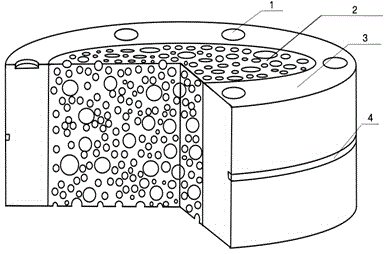
Intervertebral disc imitating spine fuser and preparing method thereof
A spinal fusion and intervertebral disc imitation technology, applied in the direction of fixator, internal fixator, internal bone synthesis, etc., can solve the problems such as difficulty in forming hydroxyapatite into a scaffold, difficulty in forming a cellulose scaffold, and difficulty in controlling the degradation rate, etc. Good clinical use effect, convenient operation, and the effect of reducing the pain of operation
- Summary
- Abstract
- Description
- Claims
- Application Information
AI Technical Summary
Problems solved by technology
Method used
Image
Examples
example 1
[0021] 1) Weigh 9 grams of chitosan powder, add it to 300 milliliters of 2% acetic acid aqueous solution, and stir mechanically for 2 hours to obtain a uniform and transparent chitosan solution. Pour it into a ring-shaped mold, immerse in a 3% sodium hydroxide aqueous solution for 2 hours to gel, wash and dry to obtain a ring-shaped cylindrical stent;
[0022] 2) Weigh 1g of chitosan and dissolve it in 100g of 2% acetic acid aqueous solution, prepare a 1% chitosan acetic acid solution, fill it into the inner cylinder of the annular cylindrical stent, and gel it with 3% sodium hydroxide melted, washed with water, and freeze-dried at -20°C to obtain a chitosan porous structure core layer with a porosity of 95% and a pore diameter of 500 μm;
[0023] 3) Dissolve interferon and bone morphogenetic protein in PBS solution, then add nano-hydroxyapatite to obtain a mixed dispersion, so that the concentration of interferon in the mixed dispersion is 10ng / mL, and the concentration of bo...
example 2
[0026] 1) Weigh 15 grams of chitosan powder, add it to 300 ml of 3% acetic acid aqueous solution, and stir mechanically for 2 hours to obtain a uniform and transparent chitosan solution. Pour it into a ring-shaped mold, immerse in a 3% sodium hydroxide aqueous solution for 2 hours to gel, wash and dry to obtain a ring-shaped cylindrical stent;
[0027] 2) Weigh 3g of chitosan and dissolve it in 100g of 2% acetic acid aqueous solution to prepare a 3% chitosan acetic acid solution, fill it into the inner cylinder of the annular cylindrical support, and then solidify it with 4% sodium hydroxide. Gelling, washing with water, and freeze-drying at -40°C to obtain a chitosan porous structure core layer with a porosity of 90% and a pore diameter of 350 μm;
[0028] 3) Dissolve interferon and bone morphogenetic protein in PBS solution, then add nano-hydroxyapatite to obtain a mixed dispersion, so that the concentration of interferon in the mixed dispersion is 100ng / mL, and the concentr...
example 3
[0031] 1) Weigh 20 grams of chitosan powder, add it to 300 milliliters of 4% acetic acid aqueous solution, and stir mechanically for 2 hours to obtain a uniform and transparent chitosan solution. Pour it into a ring-shaped mold, immerse in a 5% sodium hydroxide aqueous solution for 2 hours to gel, wash and dry to obtain a ring-shaped cylindrical stent;
[0032] 2) Weigh 5g of chitosan and dissolve it in 100g of 3% acetic acid aqueous solution, prepare a 5% chitosan acetic acid solution, fill it into the inner cylinder of the annular cylindrical stent, and gel it with 5% sodium hydroxide melted, washed with water, frozen at -40°C, and dried to obtain a chitosan porous structure core layer with a porosity of 85% and a pore diameter of 270 μm;
[0033] 3) Dissolve interferon and bone morphogenetic protein in PBS solution, then add nano-hydroxyapatite to obtain a mixed dispersion, so that the concentration of interferon in the mixed dispersion is 300ng / mL, and the concentration of...
PUM
| Property | Measurement | Unit |
|---|---|---|
| Aperture | aaaaa | aaaaa |
| Concentration | aaaaa | aaaaa |
| Aperture | aaaaa | aaaaa |
Abstract
Description
Claims
Application Information
 - R&D
- Intellectual Property
- Life Sciences
- Materials
- Tech Scout
- Unparalleled Data Quality
- Higher Quality Content
- 60% Fewer Hallucinations
Browse by: Latest US Patents, China's latest patents, Technical Efficacy Thesaurus, Application Domain, Technology Topic, Popular Technical Reports.
© 2025 PatSnap. All rights reserved.Legal|Privacy policy|Modern Slavery Act Transparency Statement|Sitemap|About US| Contact US: help@patsnap.com

