
A method for real-time monitoring of dynamic changes in the extracellular space of animal brain tissue

A technology of real-time monitoring and extracellular space, applied in the field of neurophysiological medicine, can solve the problems of large errors, influence, and inability to correctly monitor the dynamic changes of the extracellular space of the brain, and achieve the effect of enhancing physiological stress response

Inactive Publication Date: 2017-12-01
SHANTOU UNIV
View PDF0 Cites 0 Cited by
  • Summary
  • Abstract
  • Description
  • Claims
  • Application Information

AI Technical Summary

Problems solved by technology

Since this method is implemented under the assumption of a constant extracellular structure, it cannot properly monitor the dynamics of the brain's extracellular space in acute pathological conditions and provide sufficient temporal resolution
Another commonly used method in experiments is to use the reciprocal change of ion probe concentration to represent the percentage of extracellular space change, but this method has a large error and is easily affected by the perfusate in the case of brain slice perfusion
So far, there are no feasible methods to study the dynamic structure of the extracellular tissue of the brain

Method used


Image

  • A method for real-time monitoring of dynamic changes in the extracellular space of animal brain tissue
  • A method for real-time monitoring of dynamic changes in the extracellular space of animal brain tissue
  • A method for real-time monitoring of dynamic changes in the extracellular space of animal brain tissue

Examples


Embodiment 1

[0035] A computer model is used to verify the feasibility and accuracy of the phase shift method.

[0036] 1. If image 3 As shown, α(z,t) and λ(z,t) constructed in computer simulation are not only functions of time but also functions of one-dimensional space z.

[0037] 2. If image 3 As shown, the change of α(z,t) and λ(z,t) simulates a pathological state: Spreading Depression (Spreading Depression, a common pathological state), and it is assumed that the spreading depression is one-dimensional propagation, and the propagation speed u is 2.5mm / min, the time-space function graph of propagation is as follows image 3 Ψ(ξ) in 7, the simulated Ψ(ξ) function includes a square wave representing a sharp change in the extracellular structure and a sine wave with a soft change.

[0038] 3. The setting of different boundary conditions in the model can simulate the diffusion experiment in the extracellular space in body tissue (without diffusion boundary) or brain slice (with diffus...

Embodiment 2

[0046] Real-time monitoring of extracellular structural changes in the cortex of live-anesthetized rats during the onset of propagating inhibition.

[0047] 1. Sprague-Dawley adult rats (200-300 grams in weight) were intraperitoneally anesthetized with chloral hydrate, fixed their heads on a stereotaxic device, and opened a window with a diameter of 5 cm above the body motor cortex with a skull drill.

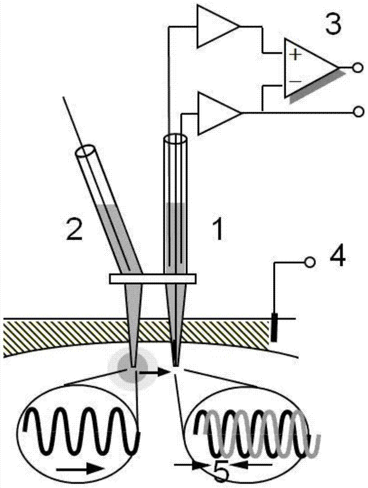
[0048] 2. Insert the potassium ion selective electrode, the ion probe (tetramethylammonium ion) release electrode-ion probe detection electrode array, and the reference electrode at 1 cm below the cortex with a micromanipulator.

[0049] 3. Select a specific frequency ω (frequency range is 0.04 ~ 0.25Hz) and phase difference ε, start the ion electrophoresis instrument according to the formula (1) and start synchronous ion probe extracellular concentration and field potential recording. After the stabilization of the standby potential, potassium ion and ion probe recordings, the...

Embodiment 3

[0060] Real-time monitoring of brain extracellular structural changes in isolated rat brain slices during the occurrence of propagating inhibition.

[0061] 1. Sprague-Dawley adult rats were intraperitoneally anesthetized with chloral hydrate (400 mg / kg), decapitated their heads, and cut their brains into 400-micron-thick slices at freezing temperatures.

[0062] 2. Place the cut brain slices in a fully immersed brain slice recording tank, perfuse with artificial cerebrospinal fluid containing 0.5mM tetramethylammonium with oxygen, and reincubate the brain slices at room temperature for 1 hour.

[0063] 3. Insert the potassium ion selective electrode, ion probe (tetramethylammonium ion) release electrode-ion probe detection electrode array, and reference electrode into the neocortex with a micromanipulator, approximately below the surface of the brain slice 200 μm, at least 1 cm away from the edge of the brain slice.

[0064] 4. Perform the phase shift method to record and in...


PUM


Abstract

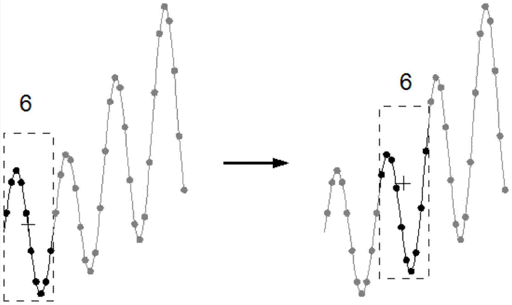
The invention discloses a method for monitoring dynamic changes of animal brain tissue extracellular gaps in real time from a frequency domain. A dynamic gap structure for monitoring brain tissues in real time is obtained by analyzing phase deviation and waveform amplitude of fixed-frequency sine diffusion waveforms of an extracellular tracing probe. Electrophoresis currents of the extracellular tracing probe comprise a constant electrophoresis current and an infinite loop sine electrophoresis current with fixed frequency changing in a sine mode along with time. According to the method, the dynamic structure changes of the living body experiment animal or in-vitro brain tissues under acute pathology states can be obtained easily and fast, and the method can be used for monitoring the dynamic changes of the brain tissue extracellular structure when the acute pathology states such as cerebral ischemia, epilepsia, transmissibility inhibition and hemorrhagic shock occur in real time, and provides the great help for learning the physiological stress reaction of brain cells and building up medical knowledge of the brain.

Description

technical field [0001] The invention belongs to the technical field of neurophysiology and medicine, and relates to a method for obtaining and processing nerve signals, in particular to a method for real-time monitoring of dynamic changes in the extracellular space of animal brain tissue. Background technique [0002] Understanding the microenvironment of the extracellular space in which brain tissue cells live is indispensable for understanding the activity of brain cells and the precursors of various diseases. The diffusion of molecules between cells is restricted by two physical factors. The first is that the narrow extracellular space (or extracellular space) makes the concentration of molecules relatively increase, and the fraction of the extracellular space in the total tissue volume is defined as α; The second is the tortuosity λ of the extracellular space. The curved path of the extracellular space makes the diffusion of the extracellular space slow. The size and st...

Claims


Application Information

Patent Timeline
Patent Type & Authority Patents(China)
IPC IPC(8): A61B5/05C12Q1/02
CPCA61B5/05
Inventor 陈致铠马霞辉
Owner SHANTOU UNIV
Learn More