LAMP1 eukaryotic expression vector having Flag label, and applications of LAMP1 eukaryotic expression vector having Flag label in lysosome separation
A eukaryotic expression vector and lysosome technology, applied in the field of molecular biology, can solve the problems that are not separated from the efficient separation of lysosomes, the specificity and titer of antibodies are not high, and the purity of lysosomes cannot be obtained. Simple operation, low cost, good separation effect
- Summary
- Abstract
- Description
- Claims
- Application Information
AI Technical Summary
Problems solved by technology
Method used
Image


Examples
Embodiment 1
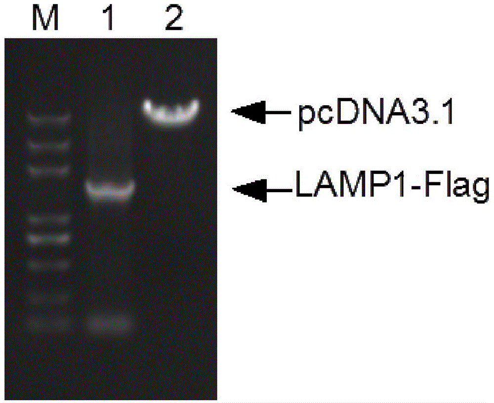
[0023] Example 1 Cloning of LAMP1-Flag Gene and Construction of pcDNA3.1-LAMP1-Flag Recombinant Plasmid
[0024] (1) RNA extraction and cDNA preparation of human cervical cancer cell Hela
[0025] will be 10 5 Hela cells were inoculated in a 6-well plate with 2 ml of DMEM complete medium containing 10% FBS, placed at 37 °C, and the volume fraction was 5% CO 2 When the cell density reaches 80%-90% confluence, treat with 1mL TRIzol, extract total RNA, and reverse transcribe into cDNA according to the instructions of Fermentas reverse transcriptase (RevertAidTM M-MuLV).
[0026](2) Cloning of LAMP1 gene and construction of pcDNA3.1(+)-LAMP2-Flag recombinant plasmid
[0027] Use the cDNA of human cervical cancer cell Hela cells as a template and the DNA sequences of SEQ ID NO.2 and SEQ ID NO.3 in Sequence Table 1 as two primers to carry out PCR amplification of the target gene, and obtain the DNA containing BamH I after purification. site and the LAMP1-Flag gene of the Xho I re...
Embodiment 2
[0030] Example 2 Establishment and screening of a cell line stably expressing LAMP1-Flag fusion protein
[0031] Human cervical cancer cells Hela were cultured in DMEM complete medium containing 10% fetal calf serum at 37°C in 5% CO 2 Cultured in medium and subcultured every 2-3 days. When the cell density in the culture dish reaches 80%-90% confluence, digest with trypsin and inoculate (0.5-1)×10 5 The cells were placed in a 6-well plate, and the next day, 2 μg of pcDNA3.1(+)-LAMP1-Flag was transfected with Lipofectamine2000 transfection reagent from Invitrogen Company. After 24 hours, the transfected cells were transferred to a 100mm culture dish, screened with 800ng / mL G418, single clones were picked out, cultured in different plates, and the single clone with the highest expression of LAMP1-Flag was screened by semi-quantitative PCR and then used 400ng / mL G418 was maintained in culture to obtain a cell line stably expressing the LAMP1-Flag fusion protein.
Embodiment 3
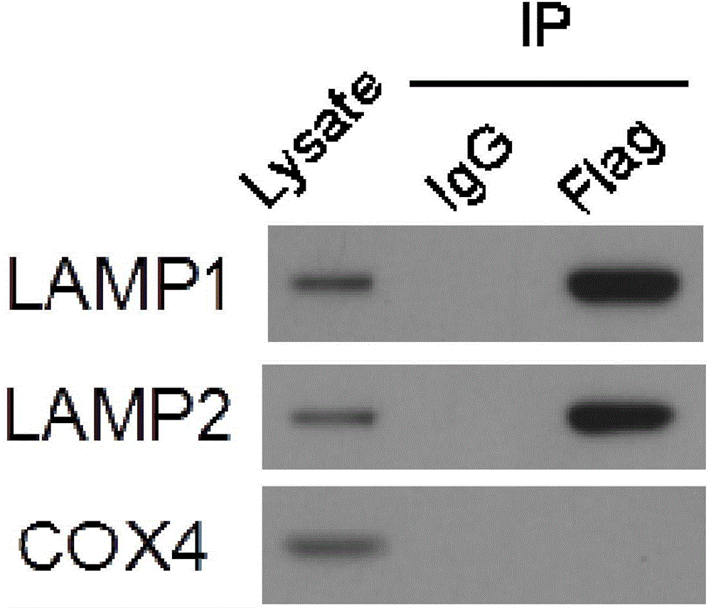
[0032] Example 3 Co-immunoprecipitation
[0033] (1) Collect Hela cells expressing LAMP1-Flag from five 100mm culture dishes by trypsinization, and use non-deformable cell lysate containing protease inhibitor PMSF (10mM HEPES, pH7.9, 1, 5mM MgCl 2 , 10mM KCl) and disrupt the cells with a homogenizer. The cell lysate was centrifuged at 1000g / min to remove cell nuclei and cell membranes, and the supernatant was recovered. The supernatant was washed with IP buffer (20mM HEPES, pH7.9, 150mM NaCl, 1.5mM MgCl 2 , 10mM KCl), add 1ug of anti-Flag M2 antibody and 25ul of protein A / G, and incubate overnight at 4°C. The precipitate was collected by centrifugation at 300g, and washed 4 times with 1ml IP buffer, and the final product obtained was the immunoenrichment product of LAMP1-Flag.
[0034] (2) Western blot detection: Add 5×protein loading buffer to the sample, put it in a 95°C water bath for 5 minutes to denature. Load 10%input, 10%anti-IgG IP, 10%anti-Flag IP. Protein sample...
PUM
 Abstract
Description
Claims
Application Information
PatSnap Eureka turns technology decisions into work you can execute. Powered by our Innovation Knowledge Graph, it runs expert workflows across engineering, life sciences, materials and intellectual property. Get your review-ready output in minutes.


