Nanometer-material-photothermal-effect-based cell detection method and cell detection device

A technology of photothermal effect and nanomaterials, applied in the direction of measuring devices, analysis materials, material excitation analysis, etc., can solve the problems of untimely diagnosis, missed breast cancer treatment time, and limitations, and achieve short detection time, improved detection sensitivity, The effect of simple detection methods

Active Publication Date: 2015-05-27
山东师大瑞柏生物科技有限公司
View PDF4 Cites 11 Cited by
  • Summary
  • Abstract
  • Description
  • Claims
  • Application Information

AI Technical Summary

Problems solved by technology

However, due to the limitations of cell detection methods, the best treatment time for breast cancer is often missed due to untimely diagnosis.

Method used


Image

  • Nanometer-material-photothermal-effect-based cell detection method and cell detection device
  • Nanometer-material-photothermal-effect-based cell detection method and cell detection device

Examples


Embodiment 1

[0039] Example 1: Preparation of MCF-7 cell detection device based on photothermal effect of nanomaterials

[0040] 1. Preparation of graphene-gold nanomaterials

[0041] Take 1 mL of 0.1% HAuCl 4 After diluting the aqueous solution to 100mL with double distilled water, transfer it to a 250mL round bottom flask and heat until the solution boils. After keeping boiling for 1 min, quickly add 3.5 mL of trisodium citrate with a mass fraction of 1% under vigorous stirring, and continue stirring until the solution turns reddish brown, then add 7 mg of graphene oxide solution until the solution turns red, and wait until the color remains stable. After the change, continue to boil and stir for 15 minutes, stop heating, and allow the solution to cool naturally. Take a certain amount of the above solution and place it in a 10mL EP tube, add 2% (m / v, unit g / mL) polyethylene glycol, ultrasonic at 211KHz for 1h, centrifuge at 8000r for 10min to remove excess graphene and polyethylene gly...

Embodiment 2

[0049] The method for MCF-7 cell detection, the steps are:

[0050] 1. Cell pretreatment: Take an appropriate amount of the cell solution to be tested and put it into a 1.5mL EP tube, centrifuge at 1500r for 5min, discard the supernatant, reconstitute with 0.01mol / L PBS and then centrifuge. Repeat the above operation three times, discard the supernatant, and dissolve the centrifugal pellet with 10 μL of 0.01mol / L PBS buffer before use;

[0051] 2. Adding samples: drop the processed MCF-7 cell solution into the sample loading area on the percolation test strip prepared in Example 1, after infiltration, wash with PBST solution three times;

[0052] 3. Add 10 μL of the graphene-gold-anti-EpCAM antibody complex prepared in Example 1, after infiltration, rinse with PBST solution three times and dry;

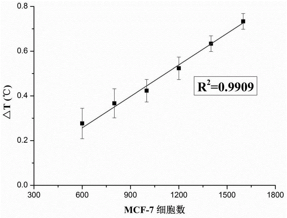
[0053] 4. Photothermal measurement: 808nm laser irradiates the sample loading area for 60s, and the infrared temperature gun records the temperature before and after and calculates t...

Embodiment 3

[0056] Example 3: Preparation of a detection device for human small cell lung cancer cells based on the photothermal effect of nanomaterials

[0057] The preparation method is the same as in Example 1, except that the antibody added during the preparation of the graphene-gold-antibody complex is 2F1 antibody.

[0058] The method of using this device to detect human small cell lung cancer cells is the same as in Example 2. The human small cell lung cancer cell solution with known cell numbers is taken for detection, and the number of cells in the human small cell lung cancer cell solution to be tested is calculated. The recovery rate is 85.7%.


PUM

PropertyMeasurementUnit
quality scoreaaaaaaaaaa

Abstract

The invention discloses a nanometer-material-photothermal-effect-based cell detection method and a cell detection device. The method comprises the following detecting steps: (1) centrifuging cell solution to be detected, discharging supernatant, redissolving with PBS (Phosphate buffer solution) and centrifuging again, repeating the operations, discharging supernatant, and dissolving centrifuged precipitates with 10mul of PBS buffer solution; (2) dropwise adding the treated cell sample onto a sample adding area of an infiltration test strip, and washing for three times with PBST (Phosphate buffer solution-tween) solution; (3) adding with 5-20muL of grapheme-gold-antibody compound into the sample adding area of the infiltration test strip, washing for three times with PBST solution and airing; and (4) radiating the sample adding area of the detection device with laser with the excitation wavelength of 808nm for 10-90s, recording the temperature before and after radiation, calculating the temperature difference, and drawing a calibration curve by use of the cell number as an x-coordinate and the temperature difference as a y-coordinate, wherien when one sample is detected, the temperature difference of the sample is detected and is substituted into the calibration curve to calculate the cell number. The detection device and the detection method have the advantages that the detection method is simple, the detection time is short, the detection sensitivity is high and the like.

Description

technical field [0001] The invention relates to a cell detection method and device based on the photothermal effect of nanometer materials. Background technique [0002] Currently, there are various methods for cell detection, such as immunocytochemical methods, PCR techniques, electrochemical methods, and microfluidic devices. Although these methods have high sensitivity and are widely used in laboratory testing and clinical diagnosis in hospitals, most methods require advanced equipment and complex operating procedures, are time-consuming and labor-intensive, and are not suitable for rapid detection. Therefore, simple and rapid detection methods still need further research. [0003] The test strip detection method is a convenient and fast detection method, including two operation modes of immunochromatography and immunofiltration. Although this method is simple and fast, its sensitivity is still low compared to other methods. In recent years, researchers have devoted th...

Claims


Application Information

Patent Timeline
Patent Type & Authority Applications(China)
IPC IPC(8): G01N21/63G01N33/574
Inventor 张鸿雁郑艳苹刘静静周建华
Owner 山东师大瑞柏生物科技有限公司
Why Eureka
Try Eureka