Fusion protein and preparation method thereof

A fusion protein and green fluorescent protein technology, applied in chemical instruments and methods, hybrid peptides, pharmaceutical formulations, etc., can solve problems such as complex and difficult methods, loss of fluorescence, and limited applications, and achieve simple and convenient operation and strong biocompatibility , The effect of simple preparation method

Inactive Publication Date: 2015-07-29
UNIV OF SCI & TECH OF CHINA
View PDF3 Cites 0 Cited by
  • Summary
  • Abstract
  • Description
  • Claims
  • Application Information

AI Technical Summary

Problems solved by technology

However, these chemical synthesis methods are usually complicated and difficult, and a specific reaction process needs to be designed for each different imaging phantom, which limits its wide application
Moreover, the biocompatibility of probes prepared by chemical methods is difficult to predict, and some have high toxicity and potential pollution hazards, which limits the application of this method in biology and medicine.
In addition, quantum dots or fluorescent dyes are prone to photobleaching and lose fluorescence, resulting in poor stability.

Method used


Image

  • Fusion protein and preparation method thereof
  • Fusion protein and preparation method thereof
  • Fusion protein and preparation method thereof

Examples


preparation example Construction

[0074] The present invention also provides a method for preparing the fusion protein of the present invention, comprising:

[0075] Step 1: obtaining a DNA molecule encoding a superpositively charged green fluorescent protein, obtaining a DNA molecule encoding a linker and a lanthanide ion-binding peptide;

[0076] Step 2: linking the DNA molecule encoding the superpositively charged green fluorescent protein with the DNA molecule encoding the linker and the lanthanide ion-binding peptide, and fusing the expression vector to construct a recombinant expression vector;

[0077] Step 3: Transform the host cell with the recombinant expression vector, induce the host cell containing the recombinant expression vector to express the fusion protein, and isolate and purify the expressed fusion protein.

[0078] At present, there are many methods for obtaining DNA molecules using genetic engineering techniques, including using restriction endonucleases to digest vectors with DNA molecul...

Embodiment 1

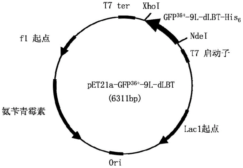
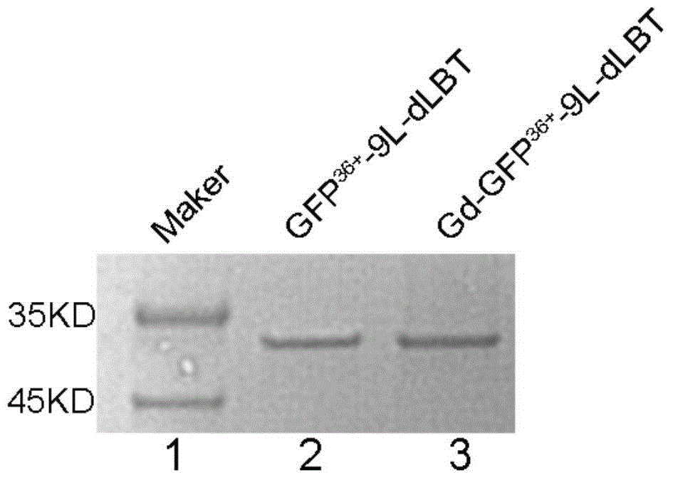
[0093] Example 1: pET21a-GFP 36+ Construction of -9L-dLBT expression vector

[0094] (1) Coding GFP 36+ The DNA sequence (SEQ ID NO: 10) was purchased from Sangong Synthesis Center. Then use this as a template to amplify GFP using primers GFP36N (SEQ ID NO:11) and GFP36C (SEQ ID NO:12) 36+ sequence, and then digested with HindIII to obtain GFP 36+ . Wherein, the primer sequence is as follows:

[0095] GFP36N (SEQ ID NO: 11):

[0096] 5'-GAACATATGGCTTCTAAAGGTGAACGCCTGTTC-3';

[0097] GFP36C (SEQ ID NO: 12):

[0098] 5'-GAAAAGCTTTTTGTAACGTTCGTCGCGGCC-3'.

[0099] (2) The DNA sequence (SEQ ID NO: 13) encoding the 27-amino acid linker and metal ion-binding peptide was purchased from Sangong Synthesis Center. Then using this as a template, primers dLBTN (SEQ ID NO: 14) and dLBTC (SEQ ID NO: 15) were used to amplify the 9L-dLBT sequence, and then digested with HindIII to obtain 9L-dLBT. Among them, the sequence is as follows:

[0100] SEQ ID NO: 13:

[0101] ATGGGTGGCTCT...

Embodiment 2

[0108] Example 2: pET21a-GFP 48+ Construction of -12L-dLBT expression vector

[0109] encodeGFP 48+ The DNA sequence (SEQ ID NO: 16) was purchased from Sangong Synthesis Center. Then use this as a template to amplify GFP using primers GFP48N (SEQ ID NO: 17) and GFP48C (SEQ ID NO: 18) 48+ Fragment, others are identical with embodiment 1. The constructed expression vector pET21a-GFP 48+ -12L-dLBT for expression of GFP 48+ -12L-dLBT protein, the sequence of its linker is (GGS) 12 . Wherein, the primer sequence is as follows:

[0110] GFP48N (SEQ ID NO: 17):

[0111] 5'-GAACAT ATGGCCAGCA AAGGCAAACG-3';

[0112] GFP48C (SEQ ID NO: 18):

[0113] 5'-GAAAAGCCTTTTTCGCCGCTTCCGCCC-3'.


PUM


Abstract

The invention relates to fusion protein which comprises super positive charge green fluorescent protein and lanthanide ion combining peptide, wherein the super positive charge green fluorescent protein is used as an optical imaging die body, and the lanthanide ion combining peptide combined with gadolinium ions is used as a paramagnetic probe. No chemical modification is needed in a preparation process, the prepared fusion protein has the advantages of strong biocompatibility, stable property and strong radiography effect and is a multifunctional intracellular imaging reagent which integrates fluorescence imaging, magnetic resonance imaging, cell penetrating and passive tumor targeting, tumor tissues of living animals can be passively targeted, tumors can be specifically imaged, any targeting die body does not need to be modified, and the fusion protein can be used as the intracellular imaging reagent, so as to realize fluorescence and magnetic resonance dual-imaging on tumor cells. The fusion protein has the advantages of simple preparation method and low cost and is suitable for industrial production.

Description

technical field [0001] The invention belongs to the technical field of biomedicine, and specifically relates to a fusion protein and a preparation method thereof, in particular to a fusion protein that can be used for fluorescence and magnetic resonance dual imaging of cells and tissues and a preparation method thereof. Background technique [0002] With the development of modern science, people's research on the essence of life activities is rapidly reducing to the level of cells and sub-cells. Molecular imaging technology provides a powerful tool for the detection of various small molecules and the tracing of metabolic pathways in cells. Imaging techniques, including optical imaging (OI), magnetic resonance imaging (MRI), positron emission tomography (PET), and computed tomography (CI), can realize real-time dynamic and seamless observation of physiological and pathological changes in biological organisms at the molecular level. The three-dimensional imaging of trauma pro...

Claims


Application Information

Patent Timeline
Patent Type & Authority Applications(China)
IPC IPC(8): C07K19/00C12N15/62C12N15/63A61K49/00A61K49/14
Inventor 吴芩刘扬中
Owner UNIV OF SCI & TECH OF CHINA
Why Eureka
Try Eureka