
Detection device for micro-fluidic biologic chip and preparation method of detection device

A technology for detection devices and biochips, which is applied in biological testing, chemiluminescence/bioluminescence, material inspection products, etc., can solve the problems of destroying biomolecules, limited application occasions, and unavoidable influence, and achieves simple preparation process and high production efficiency. The effect of simple process and stable quality

Active Publication Date: 2015-09-09
BEIJING NANO ACE TECH CO LTD
View PDF12 Cites 3 Cited by
  • Summary
  • Abstract
  • Description
  • Claims
  • Application Information

AI Technical Summary

Problems solved by technology

[0006] Patent CN 101526520B discloses a detection method and device for biological samples. Although the detection of multiple indicators of multiple samples can be realized, the biomolecules are coated on the bottom plate. Whether it is sample preparation or detection, the first plate and the second plate Neither of the two plates can move relative to each other, otherwise the biomolecules on the second plate will be destroyed. Therefore, the requirements for the experiment are strict, and it is difficult to guarantee the quality of the test.
In addition, when the device is used for sample preparation and testing, since the first flat plate has to be in motion, the position needs to be manually moved, which makes it difficult to keep the same position for sample preparation and testing, which makes it difficult to use the instrument to automatically check the test results during testing. for accurate reading
In addition, because the first plate has to be in motion and cannot be fixed, its application is limited, and it is only suitable for the temporary detection of a small number of samples in the laboratory, and cannot be used in industrial mass production; at the same time, because the first plate needs to be in the active state, it is difficult to achieve a sealed combination with the second plate, which makes it difficult to eliminate the cross-effect (contamination) of the sample liquid during sample preparation or detection, which affects the stability of the test results; and because the first plate needs to be in an active state, it is difficult to Avoid external influences on its prepared samples, so the detection conditions are strict, and it is required to be completed quickly in a short period of time from sample preparation to detection, so as to reduce the external influence on the stability of the test results

Method used


Image

  • Detection device for micro-fluidic biologic chip and preparation method of detection device
  • Detection device for micro-fluidic biologic chip and preparation method of detection device
  • Detection device for micro-fluidic biologic chip and preparation method of detection device

Examples


Embodiment 1

[0057] Embodiment 1-chip preparation

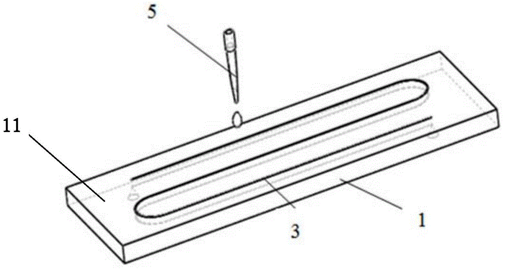
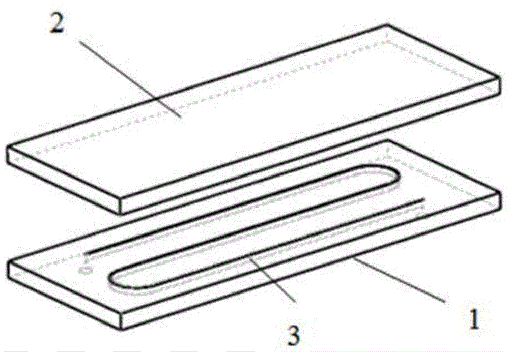
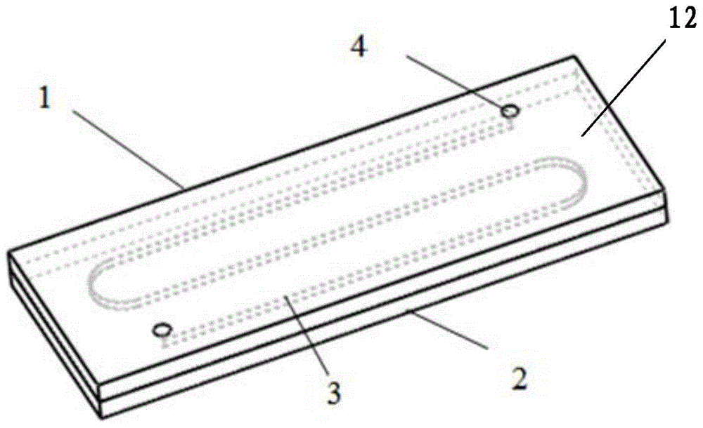
[0058] Take a polystyrene plate, and use injection molding to describe an S-shaped groove with a width of 1000 μm on one side, and the depth of the groove is 200 μm, so that the preparation of the grooved plate is completed, and a notch is formed at the corresponding position on the other side; Good AFP (alpha-fetoprotein) antibody, CEA (carcinoembryonic antigen) antibody, CA125 (sugar chain antigen 125) antibody spotting solution (sample solution for short) (the concentrations of AFP antibody, CEA antibody, and CA125 antibody are all 20 μg / ml), use the Biodot XYZ3060 spotting instrument to spot samples in the grooves of the grooved plate from left to right, the specific spotting parameters are: the point spacing is 2mm, the droplet volume is 50nl, and the distance between the needle tip and the spotting plate is within 2mm , the sampling time is controlled at 0.6-0.9ms, and the room temperature is left open for about 1-2 hours after sam...

Embodiment 2

[0059] Embodiment 2-chip preparation

[0060] Take the polydimethylsiloxane plate, and use mechanical processing to carve an S-shaped groove with a width of 100 μm on one side, wherein the depth of the groove is 400 μm, and form a notch at the corresponding position on the other side, so that the groove The plate preparation is completed; configure the spotting solution of hepatitis B surface antibody, hepatitis A surface antibody, and hepatitis C surface antibody (wherein the concentrations of hepatitis B surface antibody, hepatitis A surface antibody, and hepatitis C surface antibody are all 30 μg / ml), use Biodot XYZ3060 sample spotting instrument from Spot samples in the grooves of the grooved plate in sequence from left to right. The specific spotting parameters are: the spot spacing is 2mm, the drop volume is 50nl, the distance between the needle tip and the spotting plate is within 2mm, and the spotting time is controlled at 0.6-0.9ms , after spotting the sample, leave i...

Embodiment 3

[0061] Embodiment 3-chip preparation

[0062] Take a plexiglass plate, and use soft etching to carve a curved groove with a width of 5000 μm on one side, where the depth of the groove is 800 μm, and form a notch at the corresponding position on the other side, so that the preparation of the grooved plate is completed; configuration Good AFP antibody, CEA antibody, PSA (prostate specific antigen) antibody, CA125 sugar chain antigen antibody, CA19-9 sugar chain antigen antibody and CA15-3 sugar chain antigen antibody (AFP antibody, CEA antibody, PSA antibody , CA125 sugar chain antigen antibody, CA19-9 sugar chain antigen antibody and CA15-3 sugar chain antigen antibody concentration are all 30μg / ml), use a pipette to pipette the prepared spotting solution in the groove from left to right Spotting in the groove of the plate, the specific spotting parameters are point spacing of 2mm, droplet volume of 500nl, contact spotting, after spotting, leave it at room temperature for about...


PUM

PropertyMeasurementUnit
widthaaaaaaaaaa
depthaaaaaaaaaa
diameteraaaaaaaaaa

Abstract

The invention discloses a detection device for a micro-fluidic biologic chip and a preparation method of the detection device. The detection device mainly comprises a groove plate, a bottom plate, a groove and a notch, wherein detection points are arranged in the groove. The detection device is easy to prepare and use, and is suitable for large-scale production and application, and the detection result is stable and reliable.

Description

technical field [0001] The invention relates to a microfluidic biochip detection device and a preparation method. Background technique [0002] At present, in vitro diagnostic product technologies on the market mainly include western blotting and enzyme-linked immunodiagnostic technology (ELISA), which detect the existence of corresponding antigens or antibodies through the interaction between antigens and antibodies, so as to achieve the purpose of diagnosing a certain disease. [0003] Western blotting is a protein identification technique widely used in the field of life sciences. The test requires steps such as electrophoresis, transfer, and detection. The operation steps are complicated, and only one serum can be used at a time to prevent contamination and interfere with the test. As a result, the device is prone to false negative results. [0004] ELISA is the most widely used immunoassay technique, which uses orifice plates as solid phase carriers to measure antigens...

Claims


Application Information

Patent Timeline
Patent Type & Authority Applications(China)
IPC IPC(8): G01N21/76G01N21/78G01N33/50
Inventor 蓝海蓝洋张洪涛杨文兴时圣涛任方萍
Owner BEIJING NANO ACE TECH CO LTD
Learn More