Method for rendering tissue transparent, reagent for rendering tissue transparent, and tissue observation method

A transparent and organized technology, applied in biochemical equipment and methods, preparation of test samples, instruments, etc., can solve problems such as laborious and difficult to obtain fluorescent images, and achieve the effect of simple operation

Active Publication Date: 2015-09-30
JAPAN SCI & TECH CORP
View PDF10 Cites 12 Cited by
  • Summary
  • Abstract
  • Description
  • Claims
  • Application Information

AI Technical Summary

Problems solved by technology

[0005] In the case of three-dimensional observation of tissue structures such as neural circuits, the above-mentioned mechanical methods require the preparation of multiple consecutive tissue sections and the accumulation of these fluorescent images, which is very laborious
On the other hand, in the optical method, since light is scattered inside the organ, there is a problem that it becomes more and more difficult to obtain a fluorescence image as the depth of the part to be observed from the surface of the organ increases (observation depth limit)

Method used


Image

  • Method for rendering tissue transparent, reagent for rendering tissue transparent, and tissue observation method
  • Method for rendering tissue transparent, reagent for rendering tissue transparent, and tissue observation method
  • Method for rendering tissue transparent, reagent for rendering tissue transparent, and tissue observation method

Examples


Embodiment 1

[0112]

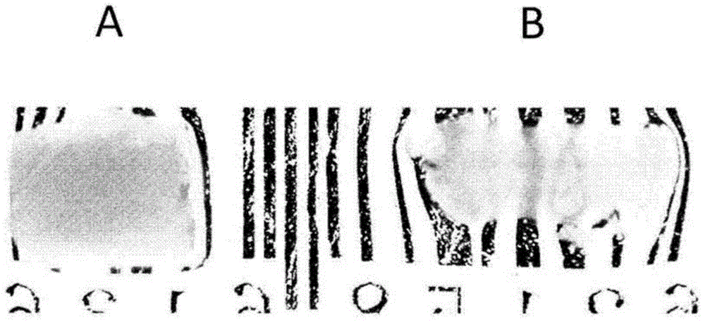
[0113] After perfusion and fixation with 4% paraformaldehyde buffer solution, the spinal cord of the rat was excised, and then immersed in the solution for 24 hours for fixation. The fixed spinal cord (diameter 3mm) was successively placed in each pretreatment solution of thiobisdiol:glycerol:sucrose=20:40:40 solution, 50:40:10 solution, and 70:25:5 solution respectively. After 24 hours of immersion, immerse in the final solution of 90:5:5 for 24 hours to perform clearing.

[0114] The result is as figure 1 Shown in B. In addition, A shows the result based on the Scale method described in Non-Patent Document 2. The tissue clearing method (B) according to the present invention can clear the spinal cord more transparently than the Scale method (A). In addition, the Scale method (A) had the problem of doubling the size of the spinal cord, whereas the tissue clearing method (B) according to the present invention did not cause such a problem.

Embodiment 2

[0115]

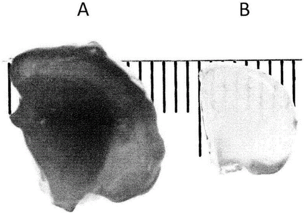
[0116] Rat brain (tissue thickness 6 mm) was fixed and cleared by the procedure described in Example 1.

[0117] The result is as figure 2 Shown in B. In addition, A shows the result based on the Scale method described in Non-Patent Document 2. The tissue clearing method (B) according to the present invention can clear the brain more transparently than the Scale method (A). In addition, the Scale method (A) had the problem that the brain swelled significantly and easily collapsed when pressed with a finger, whereas the tissue clearing method (B) according to the present invention did not cause swelling or weakening.

Embodiment 3

[0118]

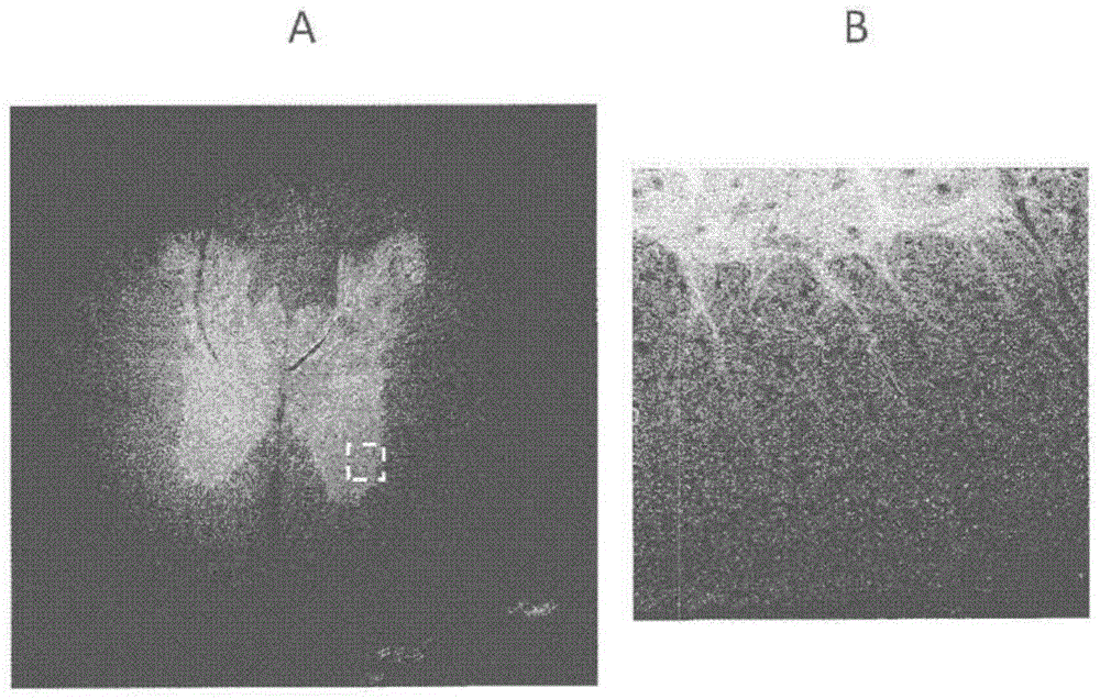
[0119] A transgenic rat expressing the fluorescent protein VENUS in the axons was prepared. The preparation of transgenic rats was carried out by the method described in Non-Patent Document 3 ("Visual properties of transgenic rats harboring the channelrhodopsin-2 gene regulated by the thy-1.2 promoter."PLoS ONE, 2009, Vol.4, No.11, e7679) conduct. The spinal cord fixed and cleared by the procedure described in Example 1 was observed with a confocal microscope (Zeiss, LSA-700).

[0120] Fluorescent images obtained as image 3 As shown in A. B is a magnified image of the area enclosed by the dashed line in A. With the tissue observation method according to the present invention, it is possible to observe nerve axons with high precision. In addition, even under the condition of relatively high concentration of thiobisethanol (90% by volume), the disappearance or attenuation of the fluorescent signal of the fluorescent protein can be suppressed.


PUM

PropertyMeasurementUnit
thicknessaaaaaaaaaa
refractive indexaaaaaaaaaa

Abstract

A method for rendering tissue transparent, said method including a step in which tissue is immersed in a water-soluble solvent including 2,2'-thiodiethanol, and glycerol and / or a non-ionic organic iodine compound, is provided as a technique with which, using a simple operation, and without using toxic or hazardous organic solvents, various organs can be rendered sufficiently transparent without inducing changes therein. In this method for rendering tissue transparent, it is preferable that a mixed solvent of 2,2'-thiodiethanol, glycerol, and a non-ionic organic iodine-compound aqueous solution be used for the water-soluble solvent.

Description

[0001] Technical division [0002] The invention relates to a tissue clearing method, a tissue clearing reagent and a tissue observation method. More specifically, the present invention relates to a tissue clearing method and the like that can easily and safely clear a tissue without causing changes in the tissue. Background technique [0003] In recent years, with the advancement of transformation technology and gene transfer technology, in various organs, only specific cells are labeled with fluorescent proteins and observed. For example, an attempt has been made to selectively fluorescently label various neural circuits in the brain, and to visualize the neural circuits in three dimensions using fluorescence as an indicator to reconstruct them. [0004] Conventionally, observation of internal tissues of organs has been performed by mechanically thinning fixed and embedded organs (or organ slices) to prepare tissue sections, and observing them under an optical microscope. ...

Claims


Application Information

Patent Timeline
Patent Type & Authority Applications(China)
IPC IPC(8): G01N1/30C12Q1/00G01N1/28G01N21/64G02B21/34
CPCG01N1/30G01N33/5082
Inventor 小野寺宏
Owner JAPAN SCI & TECH CORP
Why Eureka
Try Eureka